
Mechanism of Sanshi decoction inhibits macrophage pyroptosis by inhibiting BRD4/NF-κB/ NLRP3 pathway in the treatment of gouty arthritis
2023-03-06 09:56PIAOYongzhuQIMingmingNIEShuanglianPANGuoxiongZHANGHaoWANGXinbo
Journal of Hainan Medical College 2023年24期
PIAO Yong-zhu, QI Ming-ming, NIE Shuang-lian, PAN Guo-xiong, ZHANG Hao,WANG Xin-bo?
1. The First Affiliated Hospital of Heilongjiang University of Tradit Chin Med, Harbin 150040, China
2. Heilongjiang University of Chinese Medicine, Harbin 150040, China)
Keywords:
ABSTRACT Objective: To observe the effect of Sanshi decoction on BRD4/NF-κB/NLRP3 pathwaymediated macrophage pyroptosis, so as to elucidate the molecular mechanism of Sanshi decoction in the treatment of gouty arthritis.Methods: THP-1 was induced into macrophages with foboside and the divided into the control group, model group, low-dose, medium-dose,high-dose group of Sanshi decoction, and BRD4 inhibitor group.Except for the control group, the remaining groups were induced with monosodium urate crystals to construct a gouty arthritis cell model.The activity of macrophages was detected by CCK8, the level of macrophage pyroptosis was detected by flow cytometry, the activity of LDH, the content of IL-1β and IL-18 were detected by enzyme-linked immunosorbent assay, and the expression of related proteins in the BRD4/NF-κB/NLRP3 pathway was detected by Western blot.Results: Compared with the control group, macrophage activity was decreased in the model group, and the level of pyroptosis, LDH activity, contents of IL-1β and IL-18, expression levels of BRD4, p-NF-kB p65, NLRP3, Caspase-1 p20, and IL-1β protein were significantly up-regulated, the differences were statistically significant (P<0.05 and P <0.01).Compared with the model group, macrophage activity was up-regulated in the Sanshi Decoction, and the level of pyroptosis, LDH activity, IL-1β and IL-18 contents, expression levels of BRD4,p-NF-kB p65, NLRP3, Caspase-1 p20, and IL-1β protein were significantly decreased with statistically significant differences (P<0.05 and P <0.01).Conclusion: Sanshi decoction inhibits macrophage pyroptosis by inhibiting BRD4/NF-κB/NLRP3 pathway activation, thus improving the inflammation level of gouty arthritis.
1.Introduction
Gouty Arthritis (GA) is an inflammatory disease of the joints caused mainly by abnormal uric acid metabolism resulting in large deposits of uric acid in tissues such as the joint capsule, bursa,bone and cartilage, which in turn causes pathological damage and inflammatory reactions, and the disease is directly related to hyperuricemia[1].Clinical features such as redness, swelling, heat,and pain are often observed in the diseased tissues, causing tissue dysfunction, and greatly affecting the patient’s quality of life.The incidence of gouty arthritis has been steadily increasing in recent years and shows obvious gender and regional differences, with the prevalence of men being about three times that of women and the prevalence of people in coastal areas being higher than that in inland areas[2].Currently, there is no specific drug for GA in Western medicine, and drug treatment mainly focuses on analgesia and uric acid reduction, including glucocorticoids, non-steroidal anti-inflammatory drugs, colchicine, and other types of drugs,but such drugs can cause clear gastrointestinal damage and liver and kidney damage when taken for a long time.The characteristic diagnosis and treatment of TCM based on TCM theory have the advantages of multi-targets, side effects, and low toxicity and side effects.Studies have proved that TCM not only has pharmacological effects such as anti-inflammatory, analgesic, and antioxidant effects,but also can treat gouty arthritis by reducing the production of uric acid, promoting the excretion of uric acid, and improving the inflammation of the joints in several ways[3-4].
Macrophages (Mφ), a class of immune cells, play a very important role in the pathogenesis of gouty arthritis[5-7].Cellular pyroptosis participates in various inflammatory diseases, in which the activation of NLRP3 inflammatory vesicles is involved in the onset and development of GA through inflammatory factors such as IL-1β and IL-18[8-9].The previous study of the group confirmed the positive efficacy of San Shi Tang in the treatment group of gouty arthritis[10].Therefore, in this study, we analyzed the BRD4/NF-κB/NLRP3 signaling pathway-mediated macrophage focal death, so as to clarify the role of bromodomain-containing protein 4 (BRD4)in the regulation of macrophage focal death and to elucidate the molecular mechanism of the inhibition of gouty arthritis by Sanshi decoction.
2.Materials and methods
2.1 Experimental Animals and Cells
Forty male SD rats (6-7 weeks old) were purchased from Zhuhai Biotest Biotechnology Co., Ltd [Animal Production License No.SCXK (Guangdong) 2020-0051] and were housed in an environment with a room temperature of 20~25 ℃, circulating light, and free drinking water.
2.2 Reagents and Instrumentation
Sanshi decoction (talc 30 g, gypsum 30 g, chrysocolla 15 g, almond 15 g, zhuju 15 g, ginkgo biloba 15 g, white Tongzhi 10 g) was purchased from the Herbal Medicine Bureau of the First Affiliated Hospital of Heilongjiang University of Tradit Chin Med and was processed and prepared as a concentrate for spare use.Fobol ester was purchased from Ebixin Shanghai Biotechnology Co., Ltd (item no.abs9107); Monosodium Urate (MSU) crystals were purchased from Sigma (item no.U2875, molecular formula: C5H3N4O3Na,molecular weight 190.09), and the BRD4 inhibitor, XMD8-92, was purchased from the U.S.MedChemExpress (Item No.HY14443,molecular formula: C25H30N6O3, molecular weight 474.55).RPMI-1640 culture medium was purchased from Gibco (Item No.C11875500BT); FLICA 660-YVAD-FMK far-red caspase-1 flowthrough Immunochemistry Technologies (Item No.9122); CCK8 kit and human lactate dehydrogenase (LDH) kit were purchased from Jiangsu Enzyme Immunity Industry Co.Ltd.(item numbers KE00021 and KE00193, respectively); BRD4, NLRP3, ASC, and IL-1β primary antibodies were purchased from Wuhan Three Eagles Bio-technology Co.kB p65, p-NF-kB p65 and Caspase-1 primary antibody and HRP-labeled sheep anti-rabbit IgG secondary antibody were purchased from Abcam, USA (item numbers ab32536,ab76302, ab207802 and ab6721, respectively); β-actin primary antibody was purchased from Merck, Germany (item number:SAB1305546).The enzyme labeling instrument, electrophoresis tank, and electrophoresis instrument were purchased from Bio-Rad (models PR4100, Mini-PROTEAN Tetra, and PowerPac HC,respectively); and the flow cytometer was purchased from Shanghai Jumo Medical Instrument Company Limited (model Wmini52).The constant temperature cell culture incubator was purchased from Thermo Fisher (model HERACELL150i).
2.3 Method
2.3.1 Cell-induced differentiation
THP-1 was cultured with RPMI-1640 culture medium containing 1% penicillin-streptomycin double antibody, 10% FBS, and β-mercaptoethanol, and the cells were placed in a 37 ℃ constant temperature incubator containing 5% CO2, and 1 fluid change was performed for 2-3 d.The cells were incubated with RPMI-1640 culture medium containing 1% penicillin-streptomycin double antibody, 10% FBS, and β-mercaptoethanol.After the cells were in the logarithmic growth phase, they were inoculated in 6-well plates at a 1×10⁶cells/mL density.Subsequently, 160 nmol/L of fobol ester was added to the cells to induce the differentiation of the cells to macrophages, and the success of differentiation was suggested when the cells showed an adherent state.
2.3.2 Drug-containing serum preparation
After a 1-week period of feeding the rats were randomly divided into a control group, Sanshi decoction low-dose group, Sanshi decoction medium-dose group, and Sanshi decoction high-dose group, with 10 rats in each group.The low, medium, and high dose groups were given 26.16 g/kg, 52.32 g/kg and 104.64 g/kg of diluted Sanshi decoction concentrate in appropriate proportions by gavage,2 mL/times, respectively.In the control group, an equal volume of saline was given by gavage with reference to the drug group.1 time/day for one week.After 1 week of gavage, the rats in each group were anesthetized and blood was collected from the abdominal aorta.After centrifugation for 10 min (3 500 r/min), the serum was collected, inactivated in a water bath for 30 min, filtered through a membrane, and stored for later use.
2.3.3 Cellular grouping and pharmacological interventions
Macrophages were seeded in 6-well plates with an inoculum density of 2.5×10⁵cells/mL and 900 μL per well.Macrophages were grouped according to the protocol and subjected to pharmacological interventions: (1) Control group: 900 μL of medium + 100 μL of blank serum; (2) Model group: 900 μL of medium + 100 μL of blank serum + MSU 400 μM; (3) Sanshi decoction low-dose group: THP-1+900μL culture medium+MSU 400 μM+100 μL drug-containing serum (26.16 g/kg); (4) Sanshi decoction medium-dose group: THP-1+900 μL culture medium+MSU 400 μM+100 μL drug-containing serum (52.32 g/kg); (5) Sanshi decoction high-dose group: THP-1+100 μL culture medium+100 μL drug-containing serum (52.32 g/kg); (6) BRD4 inhibitor group: THP-1 + 900 μL culture medium +100 μL blank serum + MSU 400 μM + XMD8-92 5 μM.
2.3.4 Cellular activity assay
Meanwhile the sky had grown dark, and the breeze had turned into a stiff wind, The waves rose higher. Drops of rain slapped our faces, the temperature dropped, and thunder rumbled6 in the distance. The boat rocked like a runaway7 cradle.
Macrophages were seeded in 96-well plates (density of 3×10⁴cells/mL) with 3 replicate wells in each group, and appropriate drugs were given for intervention.20 μL of CCK8 solution was added to each well at 12 h, 24 h, and 48 h.The incubation was continued for 1 h.The absorbance value (A450) of each well was detected by an enzyme marker.
2.3.5 Flow cytometry assay
Macrophages were collected at 48h, centrifuged for 5 min (1 000 r/min), the supernatant was discarded, and then resuspended in PBS,centrifuged for 5 min (1000 r/min), washed twice with PBS after centrifugation, the supernatant was discarded, and 100 μL of cells were left in the centrifugation tubes, and then resuspended by adding FAM-FLICA-labeled Caspase-1 antibody and mixing, incubated for 60 min in a 5% CO2incubator under protected light, and then washed twice with 1×Wash Buffer, and resuspended by adding PI staining solution (1:100).After incubation for 60 min at 37 ℃ and 5% CO2in an incubator protected from light, the cells were washed twice with 1×Wash Buffer and resuspended by adding PI staining solution (1:100), and then detected by flow cytometry on board, and analyzed by Flow Jo 7.6.2 software.Cells that were double positive for Caspase-1 and PI were used to represent the level of focal death[11].
2.3.6 ELISA assay
Macrophage cultures were collected at 48 h, and ELISA kits were used to detect the levels of LDH, IL-1β, and IL-18 in the supernatants of cell cultures of each group, and the experimental operation was carried out in strict accordance with the instructions of the kits.
2.3.7 Western blot detection of BRD4/NF-κB/NLRP3 pathway-related protein expression
Cells of each group were collected at 48 h, and cell lysis was performed on ice after adding RIPA.The supernatant was discarded after centrifugation for 15 min (2 000 r/min), and the concentration of protein was detected by the BCA method.On the sample, the protein was separated by SDS-PAGE and then transferred to PVDF,and the membrane was closed by adding 5% skimmed milk at room temperature for 1 h, then the primary antibody diluted in the appropriate proportion was added: BRD4, NF-kB p65, p-NF-kB p65, NLRP3, ASC, Caspase-1, IL-1β and β-actin, and then HRP was added after incubation at 4 ℃ overnight.After the addition of HRP-labeled sheep anti-rabbit IgG secondary antibody, the incubation was continued at room temperature for 2 h.A gel imaging system was used for imaging, and the relative expression level of the target proteins was calculated by the ratio of the target protein to the internal reference, using β-actin as the internal reference.
2.4 Statistical analyses
SPSS 26.0 software was used to analyze the data, ANOVA was used to compare the data between multiple groups, and LSD test was used to compare the two-by-two data between groups, and P<0.05 was used to indicate that the difference was statistically significant.
3.Results
3.1 Effect of Sanshi decoction on macrophage activity
Comparison of macrophage activity between the groups at 12 h showed that the cell activity in the model group was significantly lower than that in the control group, and the difference was statistically significant (P<0.05).Compared with the model group,the activity of macrophages in the high-dose and inhibitor groups of Sanshi decoction was significantly increased, and the differences were statistically significant (P<0.05).Comparison of macrophage activity between groups at 24 h and 48 h showed that the activity of the cells in the model group was significantly reduced compared with that in the control group, and the differences were statistically significant (P<0.01).Compared with the model group, the activity of macrophages in the low-dose, medium-dose, high-dose Sanshi decoction, and inhibitor groups were significantly increased, and the differences were all statistically significant (P<0.05 and P<0.01).See Table 1
Tab 1 Comparison of macrophage activity in each group(n=3, ±s, OD)

Tab 1 Comparison of macrophage activity in each group(n=3, ±s, OD)
Note: Compared with the control group, *P<0.05, **P<0.01; compared with the model group, ●P<0.05, ●●P<0.01.
Grouping 12h 24h 48h Control group 0.62±0.07 0.82±0.10 0.93±0.08 Model Group 0.39±0.07* 0.56±0.08** 0.61±0.08**Sanshi decoction Low Dose Group 0.42±0.04 0.62±0.04 0.72±0.03●Sanshi decoction Medium Dose Group 0.55±0.17 0.71±0.03● 0.77±0.04●●Sanshi decoction High Dose Group 0.61±0.09● 0.79±0.02●● 0.80±0.02●●Inhibitor group 0.61±0.07● 0.78±0.11●● 0.81±0.04●●F 3.606 6.009 10.963 P 0.032 0.005 0.000
3.2 Effect of Sanshi decoction on macrophage pyroptosis
Compared with the control group, the proportion of macrophage Caspase-1/PI positivity was significantly higher in the model group, and the difference was relatively statistically significant(P<0.01).Compared with the model group, macrophage activity was significantly increased in the low-dose, medium-dose, high-dose Sanshi decoction, and inhibitor groups and the differences were all statistically significant (P<0.01), with the high-dose of Sanshi Tang being the best.See Table 2 and Figure 1.
Tab 2 Comparison of macrophage pyroptosis levels in each group(n=3, ±s)

Tab 2 Comparison of macrophage pyroptosis levels in each group(n=3, ±s)
Note: Compared with the control group, **P<0.01; compared with the model group, ●●P<0.01.
Grouping Positive expression of Caspase-1(%)Control group 4.83±0.84 Model Group 17.40±1.30**Sanshi decoction Low Dose Group 14.43±0.97●●Sanshi decoction Medium Dose Group 11.30±0.56●●Sanshi decoction High Dose Group 6.64±0.42●●Inhibitor group 7.09±0.24●●F 113.202 P 0.000
3.3 Effect of Sanshi decoction on macrophage LDH, IL-1β and IL-18 content
Compared with the control group, the release of macrophage LDH and the content of IL-1β and IL-18 in the supernatant were significantly increased in the model group, and the difference was statistically significant in comparison (P<0.01).Compared with the model group, the release of macrophage LDH and the contents of IL-1β and IL-18 in the supernatant in the low-dose, medium-dose,high-dose Sanshi decoction, and inhibitor groups were significantly decreased, and the differences were statistically significant when compared (P<0.05 and P<0.01), in which the efficacy of Sanshi decoction showed a concentration-dependent manner, and it was best in the high dose of Sanshi decoction.See Table 3.
Tab 3 Comparison of LDH and inflammatory factor levels in each group (n=3, ±s)

Tab 3 Comparison of LDH and inflammatory factor levels in each group (n=3, ±s)
Note: Compared with the control group, *P<0.05, **P<0.01; compared with the model group, ●P<0.05, ●●P<0.01.
Grouping LDH(U/L) IL-1β(pg/mL) IL-18(pg/mL)Control group 102.75±7.52 8.66±1.54 10.53±1.11 Model Group 214.78±7.24** 41.99±2.44** 39.92±2.70**Sanshi decoction Low Dose Group 185.28±9.30●● 35.31±3.10●● 35.32±2.15●Sanshi decoction Medium Dose Group 174.39±7.72●● 23.72±3.39●● 29.50±1.44●●Sanshi decoction High Dose Group 142.07±3.53●● 11.13±1.89●● 20.97±1.62●●Inhibitor group 147.63±6.85●● 14.99±2.11●● 20.45±1.16●●F 88.597 92.598 P 0.000 0.000 0.000
3.4 Effect of Sanshi decoction on BRD4/NF-κB/NLRP3 pathway in macrophages

Fig 1 Comparison of macrophage pyroptosis levels among groups
Compared with the control group, the expression levels of BRD4,p-NF-kB p65, NLRP3, Caspase-1 p20 and IL-1β proteins were significantly up-regulated in macrophages in the model group, and the differences were all statistically significant (P<0.01), whereas there were no significant changes in the expression of NF-kB p65,ASC and Pro-IL-1β proteins, and the differences were all were not statistically significant (P>0.05).Compared with the model group,the expression levels of BRD4, p-NF-kB p65, NLRP3, Caspase-1 p20, and IL-1β were significantly reduced in macrophages in the low-dose, medium-dose, high-dose Sanshi decoction, and inhibitor groups, and the differences were all statistically significant (P<0.05 and P<0.01), while no significant changes were found in the expression of NF-kB p65, ASC and Pro-IL-1β proteins.1β protein expression was not significantly changed, and the differences were not statistically significant when compared (P>0.05), in which the efficacy of Sanshi Tang showed a concentration-dependent manner,with the high dose of Sanshi decoction being the best.See Table 4-5 and Figure 2.
Tab 4 Comparison of BRD4, NF-κB and NLRP3 protein expression in macrophages of each group(n=3, ±s)

Tab 4 Comparison of BRD4, NF-κB and NLRP3 protein expression in macrophages of each group(n=3, ±s)
Note: Compared with the control group, **P<0.01; compared with the model group, ●P<0.05, ●●P<0.01.
Grouping BRD4/β-actin p-p65/total-p65 NLRP3/β-actin Control group 0.14±0.08 0.24±0.03 0.11±0.05 Model Group 0.86±0.11** 0.93±0.12** 0.93±0.07**Sanshi decoction Low Dose Group 0.73±0.09 0.91±0.04 0.65±0.16●●Sanshi decoction Medium Dose Group 0.38±0.13 0.70±0.18● 0.44±0.15●●Sanshi decoction High Dose Group 0.28±0.03● 0.48±0.04●● 0.32±0.04●●Inhibitor group 0.29±0.06● 0.47±0.03●● 0.33±0.07●●F 31.096 25.671 24.140 P 0.000 0.005 0.000
Tab 5 Comparison of ASC, Caspase-1 and IL 1β protein expression in macrophages of each group(n=3, ±s)
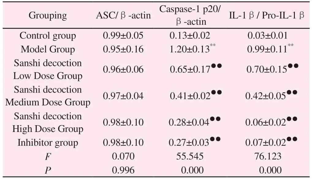
Tab 5 Comparison of ASC, Caspase-1 and IL 1β protein expression in macrophages of each group(n=3, ±s)
Note: Compared with the control group, **P<0.01; compared with the model group, ●●P<0.01.
Grouping ASC/β-actin Caspase-1 p20/β-actin IL-1β/ Pro-IL-1β Control group 0.99±0.05 0.13±0.02 0.03±0.01 Model Group 0.95±0.16 1.20±0.13** 0.99±0.11**Sanshi decoction Low Dose Group 0.96±0.06 0.65±0.17●● 0.70±0.15●●Sanshi decoction Medium Dose Group 0.97±0.04 0.41±0.02●● 0.42±0.05●●Sanshi decoction High Dose Group 0.98±0.10 0.28±0.04●● 0.06±0.02●●Inhibitor group 0.98±0.10 0.27±0.03●● 0.07±0.02●●F 0.070 55.545 76.123 P 0.996 0.000 0.000
4.Discussion

Fig 2 effect of astragalus ginseng soup on BRD4/NF-κB/NLRP3 pathway in macrophages
Macrophages are a class of immune cells with multiple functions that participate in the body’s intrinsic and adaptive immunity.They are widely distributed and can be found in a variety of tissues and organs, contributing to the maintenance of homeostasis in the body’s immune environment.Previous studies of macrophage death have focused on apoptosis or necrosis, but with the development of research in the fields of inflammatory response and pathogen infection, cellular pyroptosis has gradually entered the research field.Scorched death occurs mostly in macrophages and is a programmed death process accompanied by the release of inflammatory factors caused by intrinsic immunity[12].The results of this study showed that MSU induction was able to reduce macrophage activity,upregulate macrophage Caspase-1 positive expression, and promote macrophage pyroptosis.In contrast, high-dose Sanshi Tangcontaining serum could effectively increase macrophage activity,down-regulate macrophage Caspase-1 positive expression, and inhibit macrophage pyroptosis, with effects comparable to those of BRD4 inhibitors.
As the main secreted form of IL-1, IL-1β can induce the release of other inflammatory molecules through amplification of signaling linkage responses and, in a synergistic manner, promote inflammation.Elevated IL-1β can be observed in different clinical stages of GA, and IL-1β can be used as an observational indicator to feedback the inflammatory activity and disease severity in GA [13-14].IL-18, one of the family members of IL-1, induces the synthesis of IFN-γ, which participates in the immune response, induces immune injury, and then aggravates the inflammatory response.In GA, IL-18 induces triggers and maintains the persistence of the inflammatory response[15].LDH is an enzyme-like molecule present in the cytoplasm, which not only participates in intracellular lactate metabolism processes but also serves as a clinical indicator for assessing tissue damage and inflammatory status, among others.Studies have confirmed that increased LDH promotes the upregulation of genes such as Caspase-1, IL-1β, and GSDMD in the cellular pyroptosis pathway, thereby exacerbating the inflammatory response to promote the occurrence of cellular pyroptosis[16].The results of the present study showed that MSU induction was able to significantly increase macrophage IL-1β, IL-18, and LDH content, while high-dose Sanshi Tang-containing serum was able to effectively inhibit macrophage IL-1β, IL-18, and LDH secretion,with an effect comparable to that of BRD4 inhibitors.
BRD4 belongs to the BET family, which also includes three members, BRD2, BRD3, and BRDT.BRD4 contains two bromodomain structural domains and an interleukin structural domain, which can help BRD4 realize its regulatory roles as a genetic regulator and transcriptional cofactor.It has been demonstrated that BRD4 binds to the promoters of inflammatory cytokine genes in macrophages and directly regulates the expression of inflammatory factors and inflammatory responses in macrophages[17].A BRD4-specific inhibitor (JQ-1) is able to down-regulate the expression of key genes and inhibit the onset of cellular pyroptosis in activated macrophages[18-19].NF-κB is a major transcription factor in macrophages one of the major transcription factors in macrophages,which is involved in the regulation of immune and inflammatory responses in macrophages.Studies have confirmed that TCM can reduce the occurrence of cell death in many diseases by inhibiting the NF-κB signaling pathway[20-22].NLRP3 inflammasome is an intracellular multiprotein complex that has multiple roles such as proinflammatory, antipathogen infection, and immune regulation,and has been associated with a variety of inflammatory diseases and autoimmune disorders[23-24].Upon receiving a signaling stimulus, the NLRP3 inflammatory vesicle, consisting of NLRP3,an Apoptosis-associated speck-like protein containing a CARD(ASC) and pro-Caspase-1, is assembled and activated[25].It was demonstrated that NLRP3 may induce the release of inflammatory factors, such as IL-1β and IL-18, through the activation of GSDMD proteins, which in turn leads to macrophage pyroptosis[26].During this process, NLRP3 contributes to the formation of the ASC half-site, which cleaves and activates caspase-1, which postshears the GSDMD protein and releases its N-terminal fragment[27].This fragment binds to phosphodiester molecules on the cell membrane to form a pore, which not only contributes to the release of inflammatory factors such as IL-1β and IL-18 but also leads to the rupture and breakdown of the cell membrane, which ultimately allows cellular pyrolysis to occur[28].The activation of NLRP3 inflammatory vesicles throughout the process is critical in mediating the subsequent onset of cellular pyroptosis.
GA belongs to the scope of paralysis in the category of Tradit Chin Med (TCM), which considers wind, cold, and dampness as the causes of paralysis, and can show different symptoms according to the preference of wind, cold, and dampness.Three Stone Soup is from “Regulations on Warm Diseases”, which is composed of Flying Slippery Stone, Gypsum, Cold Water Stone, Almonds, Bamboo Roots, Silver Flower, and White Tongzhi.This formula takes talc as the ruler, taking its cold nature and sweet flavor, clearing heat, and removing evil and the function of the surface and the inside of the body.The subject medicine is used to help the ruler, and gypsum,cold water stones, silver flowers, etc.are taken for their ability to clear heat and benefit the orifices.Almonds are also used to promote the upper focus, and Bamboo Root is used as an adjuvant for the middle focus.Tongcao is used as an adjuvant.Tongcao is used to facilitate the dispersal of medicinal power in the body and is used as an enabler.The whole formula is mainly for clearing up and eliminating heat and dampness to eliminate evils.Relevant data mining and literature have shown that several drugs such as talc,gypsum, and ginkgo are commonly used in the modern treatment of GA in Sanshi decoction[29].In this study, MSU induction was found to be able to up-regulate the expression of BRD4, p-NFkB p65, NLRP3 inflammatory vesicles, and IL-1β, whereas the drug-containing serum of Sanshi decoction could effectively down-regulate the expression of BRD4, p-NF-kB p65, NLRP3 inflammatory vesicles, and IL-1β.
In summary, Sanshi decoction can improve the inflammation level of gouty arthritis by inhibiting the activation of the BRD4/NF-κB/NLRP3 pathway and thereby inhibiting macrophage pyroptosis.
 Journal of Hainan Medical College2023年24期
- Journal of Hainan Medical College的其它文章
- Meta-analysis of the impact of hyperuricemia on contrast agent-related acute kidney injury after percutaneous coronary intervention
- Research progress on the drug action and resistance mechanism in Mycobacterium tuberculosis
- Macrophage-derived exosomes mediate mir-222 targeting Caspase-10 to promote glioma proliferation
- Mechanism of action of verbascoside in mice with Parkinson's disease based on molecular docking, molecular dynamics and in vivo experiments
- Effects of female body mass index on embryo development and ART outcomes
- Prediction of potential targets and molecular mechanisms of Tripterygium hypoglaucum for oral lichen planus based on network pharmacology and molecular docking