
Establishment of extensively drug-resistant Pseudomonas aeruginosa pneumonia model in rat
2023-12-06 07:53ANRanLIUFengyunZHAOQiliangCAIWenxinSUNHongyuanCHENMinghuCHANGLiYINYanzhongLIUKaiZHANGHuiqiLIUMin
Journal of Hainan Medical College 2023年15期
AN Ran, LIU Feng-yun, ZHAO Qi-liang, CAI Wen-xin, SUN Hong-yuan, CHEN Ming-hu, CHANG Li, YIN Yan-zhong, LIU Kai, ZHANG Hui-qi?, LIU Min?
1.Department of Infectious Diseases, First Teaching Hospital of Tianjin University of Traditional Chinese Medicine, Tianjin 300193, China
2.National Clinical Research Center for Chinese Medicine Acupuncture and Moxibustion, Tianjin 300193, China
3.Tianjin University of Traditional Chinese Medicine, Tianjin 301617, China
Keywords:
ABSTRACT Objective: To establish extensively drug-resistant Pseudomonas aeruginosa (XDR-PA)infection-induced pneumonia model in rats.Methods: Twenty-four male SD rats were randomly divided into blank group, low bacterial group, medium bacterial group, and high bacterial group.The low, medium and high bacterial groups were given intratracheal instillation of 0.1 mL of bacterial suspension (bacterial concentration in turn is 7.5×10⁹, 3×10¹⁰, 6×10¹⁰ CFU/mL), while the blank group were given the same volume of sterile normal saline.After modeling, the general conditions of rats in each group were observed, including mental state,hair, respiration, activity, eating, weight, and the survival curve was drawn.The pathological characteristics of lung tissue and the infiltration of inflammatory cells were observed.Pathogenic identification of each group was carried out by bacterial culture of lung tissue homogenate.Results: The general state of the blank group was normal, and the rats in other groups showed signs of mental depression, bristling, shortness of breath, even oral and nasal bleeding, decreased food intake and activity, and significant weight loss, and different degrees of death within 48 hours, the difference was statistically significant (P<0.05).Pathological results showed that the alveolar structure of rats in the blank group was complete, and the alveolar space was clear without exudation.The lung tissue of the low and medium bacterial groups showed obvious inflammatory cell infiltration, alveolar structure destruction, alveolar septum thickening, interstitial edema, but the pathological damage of the medium group was more severe, with a mortality rate of up to 50%, and the mortality rate of the low bacterial group was 17%.In the high bacterial group, red blood cells, inflammatory cells and a large amount of fibrin-like exudation can be seen in the alveolar space, which has the pathological characteristics of acute respiratory failure, and the mortality rate is as high as 67%.The results of bacterial culture of lung tissue homogenate showed that the blank group had no bacterial colonies, while PA colony growth can be seen in low, medium and high bacterial groups.Conclusion: Intratracheal instillation of low bacterial count (0.1 mL of 7.5×10⁹ CFU/mL)XDR-PA bacterial suspension can successfully construct a rat pneumonia model of XDR-PA infection.
1.Introduction
Pseudomonas aeruginosa (P.aeruginosa, PA)is a common pathogen of hospital acquired pneumonia and ventilator-associated pneumonia[1,2].PA is characterized by easy colonization, mutation,and multi-drug resistance, and its drug resistance mechanism is complex[3], which can easily produce superbugs and cause infections that are difficult to treat.One study[4] showed that drugresistant PA infection increased the risk of death by 24% compared to patients with non-resistant PA infection, and those with multidrug resistant PA infection had more than a 2-fold increased risk of death in hospital compared to those with sensitive PA infection.Extensively drug-resistant PA (XDR-PA) is a common type of PA resistance in clinical practice, which leads to severe pneumonia, long disease duration, high mortality rate, and severely limited antibiotic choices, making clinical treatment extremely difficult and increasing the socioeconomic burden.The construction of an animal model of XDR-PA pneumonia is an important part of studying its resistance mechanism and testing the clinical efficacy of new antibiotics.In this study, we intend to further compare the lung pathological characteristics of rats with pneumonia caused by different bacterial loads (low, medium and high) on the basis of preliminary preexperiments, and finally establish the best XDR-PA pneumonia model.
2.Materials and Methods
2.1 Materials
2.1.1 Experimental animals
Male SPF-grade SD rat, weighing 160±10 g, purchased from Beijing HFK Bioscience co., Ltd.with license number SCXK(Beijing) 2019-0008.All animals were acclimatized and housed for 5 days under the following conditions: room temperature 24±1 ℃,relative humidity 55±10% and 12-hour light/dark cycle.
2.1.2 Main reagents and instruments
XDR-PA strain (210608091) was isolated and purified from clinical sputum samples by the microbiological laboratory of the Laboratory Department of First Teaching Hospital of Tianjin University of Traditional Chinese Medicine.The strain was identified as PA using matrix-assisted laser desorption ionization-time-of-flight mass spectrometry (Vitek MS system, bioMérieux), and the drug sensitivity was determined using the viteck2-compact (bioMérieux)fully automated bacterial identification drug sensitivity analyzer.The strain was sent to Beijing Meiyou Annuo Biotechnology Co.,Ltd.for sequencing analysis, and was identified as PA by 16SrDNA molecular biology, with 100% homology to Pseudomonas aeruginosa strain JCM 5962 MK796437.1.Columbia blood plate (Guangdong Huankai Microbial Sci.&Tech Co., Ltd., Lot No.: M2357Y).Eosin staining solution (Beijing Zhong Shan-Golden Bridge Biological Technology Co., Ltd., Lot No.: ZLI-9613).Hematoxylin staining solution (Beijing Zhong Shan-Golden Bridge Biological Technology Co., Ltd., Lot No.: ZLI-9610).Acid ethanol differentiation solution(Shanghai Yuanye Bio-Technology Co., Ltd., No.: R20777).
Turbidimeter (Mérieux DensiCHEK Plus), incubator (Shanghai Yiheng Technology Instrument Co., Ltd., model: DHP9121B),dual-function air bath thermostat shaker (Changzhou Jintan Xuri Experimental Instrument Factory, model: ZD-85), centrifuge (Beijing Times Beili Centrifuge Co., Ltd., model: DT5-3).Dehydrator(Leica Instrument Ltd., model: LEiCA TP-1020), pathological slicer(Leica Instrument Ltd., model: LEiCA HistoCore MULTICUT),embedding machine (Jinhua Hisure Scientific Co., Ltd., model:ES500-1), freezing table (Jinhua Hisure Scientific Co., Ltd., model:ES500-2C), tissue spreading machine (Jinhua Hisure Scientific Co., Ltd., model: ES500-3H), oven (Shanghai Yiheng Technology Instrument Co., Ltd., model: BPG-9106A), microscope (OLYMPUS,model: OLYMPUS CKX43-LP), tissue grinder (Wuhan Servicebio Technolony Co., Ltd., model: KZ-III-F), panoramic film sweeper(3DHISTECH).
2.2 Methods
2.2.1 Preparation of bacterial suspension
XDR-PA strain was taken out from -80 ℃ refrigerator and thawed at room temperature, inoculated to blood plate with disposable aseptic inoculation ring, zoned delineation to isolate single colonies,inverted to incubate and cultivate in thermostat at 37 ℃ for 18~24 h.Gray-green colonies which were round, flattened and moist, with irregular edges and a special ginger flavor were seen, surrounded by transparent hemolytic ring, and stained microscopically as gramnegative bacilli.The single colony was selected and inoculated to nutrient broth, shaking the bed at 37 ℃ overnight, speed 200 r/min.The broth was observed to be uniformly turbid.After centrifugation(2 500 r/min) for 10 min, the supernatant was removed and the bottom layer bacteria were retained, and then an appropriate amount of sterile normal saline was added to rinse and mix.The original bacteria suspension was obtained after repeating the above operations.A few volume of original bacterial solution was taken as the sample, and the concentration of the dilution solution was determined by turbidimeter detection after the multiple dilution of the sample, and then the original bacterial solution concentration was calculated by multiplying the dilution multiple.Finally, after the conversion and multiple dilution, the bacterial suspension with low,medium and high concentrations was prepared for standby: 7.5 × 10⁹CFU/ml (low), 3 × 10¹⁰CFU/ml (medium), 6 × 10¹⁰CFU/mL (high).
2.2.2 Grouping and modeling
Twenty-four SPF grade SD rats were randomly divided into four groups: blank group, low bacterial group, medium bacterial group and high bacterial group.The rats were weighed and injected intraperitoneally with sodium pentobarbital (30 mg/kg).After anesthesia, the rats were placed in a supine position on the animal operating table, and the head and limbs were fixed.The head end of the rat plate was elevated at an angle of 30°to the operating table.The neck hair was shaved, and the neck skin was exposed.The skin was disinfected locally along the tracheal route.Then, the skin and subcutaneous tissue were incised longitudinally in the middle of the anterior neck, and bluntly dissected until the trachea was exposed.After aspirating 0.1 mL of XDR-PA bacterial suspension with a 1 mL syringe, under direct vision, the needle is tilted 30°towards the direction of the lower airway and inserted between the two cartilage rings.After slowly injecting the bacterial solution, the needle was immediately pulled out.The rats were kept in an upright position for 30 s and rotated left and right to evenly distribute the bacterial solution to the lungs on both sides.After that, the muscle layer was immediately used to seal the drug perforation, the neck incision was sutured, disinfected, and then the rats were placed slightly high on the head.After awakening from anesthesia, the rats were released to return to the cage, free to move around, and given water and feed.The low, medium and high bacterial group were tracheally injected with 0.1 mL of bacterial suspension at concentrations of 7.5×10⁹CFU/mL, 3×10¹⁰CFU/mL, and 6×10¹⁰CFU/mL, with inoculum amounts of 7.5×10⁸CFU, 3×10⁹CFU, and 6×10⁹CFU, respectively;the blank group was tracheally injected with 0.1 mL of sterile saline.
2.3 Observation indicators
2.3.1 General status of rats
After modeling, we observed the general conditions of rats in each group, such as mental status, hair, respiration, activity, feeding,weight, etc.We observed whether the rats died or not, and recorded the time of death to draw the survival curve.
2.3.2 Histopathologic characteristics of the lungs
The chest cavity was opened in aseptic operation, and the pathological changes of the lung tissue in general were observed to see the size of the lung volume and the variations in color and texture, and to observe whether there were hemorrhagic spots,nodules, consolidation and other alterations on the surface of the lung tissue.The left lung tissue was taken and fixed in 4%paraformaldehyde solution for more than 72 h.After dehydration,embedding, sectioning, HE staining, the changes of lung histopathology were observed by digital pathological slice scanning imaging system.
2.3.3 Bacteriologic identification of lung tissue
The lower lobe of the right lung tissue was isolated, a certain amount of lung tissue (50~70 mg) was weighed, and 10 times the volume of sterile physiological saline was added to grind and homogenisein at low temperature.100 uL of homogenate was evenly coated on the blood plate with a disposable sterile coating rod.The coated blood plate was incubated in a 37 ℃ constant temperature incubator for 18~24 h.The growth of colonies on the blood plate was observed, and the bacteriological identification was carried out.
2.4 Identification of rat XDR-PA pneumonia model
According to the clinical manifestations of PA pneumonia rats,the pathological characteristics of lung tissue, and the results of bacteriological identification of lung tissue, combined with the evaluation opinions of major researchers, pathologists, and microbiology experts, a consensus was reached to identify that the rat XDR-PA pneumonia model was successfully constructed.
2.5 Statistical methods
Kaplan-Meier method was used for statistical analysis to compare whether the survival curves of rats in each group were consistent.P<0.05 indicated that the difference was statistically significant.
3.Results
3.1 General status of rats
Rats in the blank group were mentally active and responsive, with glossy fur and a slight weight loss on the first day after modeling,followed by a gradual increase in body weight (Fig.1, right).The rats in the low, medium and high bacterial groups were depressed and curled up on one side of the cage with erect and lustreless fur.They had sluggish response to external stimuli, reduced avoidance behaviour, bilateral chest undulation, accelerated respiratory rate,partly accompanied by respiratory murmur, bleeding from the mouth, nose and inner canthus of the eyes, reduced food intake,significant weight loss, gradual weight gain after 2~3 d, and death of varying degrees within 48 h (Fig.1, Table 1).The overall distribution of survival time was statistically different among the four groups,Logrankχ2=8.248, P<0.05.
3.2 Histopathologic observation of the lungs
Observed by the naked eye, the lung lobes of rats in the blank group were pale pink, with normal volume and no obvious congestion and edema.The lung volume, weight and tissue congestion of rats in the low, medium and high bacterial groups were increased, and scattered bleeding points could be seen.In the high bacterial group,tissue congestion and bleeding were serious, and some lung lobes were consolidated.As shown in Figure 2, microscopic observation showed that the alveolar structure of rats in the blank group was complete, the alveolar cavity was clear without exudation, and the alveolar septum was slightly thickened.In the low and medium bacterial groups, there were more inflammatory cell infiltration in the bronchiole lumen, bronchioles and around blood vessels,serous exudation and inflammatory cells could be seen in the alveolar cavity, the alveolar structure was damaged, and the foci had incomplete alveolar expansion or alveolar collapse, fusion, alveolar septum thickening, pulmonary interstitial congestion and edema.In the high bacterial group, inflammatory secretions and inflammatory cells could be seen in the bronchiole lumen, the alveolar wall was dilated and congested, there were red blood cells and a large number of cellulose like exudates in the alveolar cavity, and a large number of inflammatory cells could be seen in some alveolar cavities.
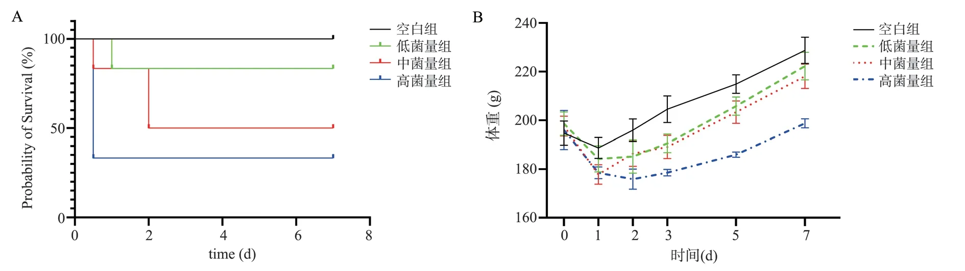
Fig 1 Survival curve and weight changes of XDR-PA pneumonia rats

Tab 1 Death of animals in each group within 48 hours of modeling
3.3 Pathogenetic identification of groups
A combination of tissue homogenization and bacterial culture was used to identify the pathogenicity of each group.As shown in Figure 3, flat, moist and metallic colonies can be seen in the homogenate culture of lung tissue in the low, medium and high bacterial groups,while no PA colony can be seen in the blank group.

Fig 2 Histopathological sections (HE staining, 200×) of the lungs of each group of rats (pre-experimental results)

Fig 3 Culture results of lung tissue homogenate in each group (preexperimental results)
4.Discussion
There is no unified standard for the establishment of animal models of PA pneumonia.According to the literature, available experimental animals include mice, rats, guinea pigs, rabbits, pigs, etc., of which the most widely used are rodents.SD rats are large in size, easy to operate, sampling, better tolerant to bacterial infections, with lower mortality rate, convenient to feed, and inexpensive[5,6].They are currently the most popular animals for the experimental study of PA pneumonia in China, therefore, they were selected for this experiment[7-10].The experimental strains used for modeling are both standard strains of Pseudomonas aeruginosa and clinical isolates,but there are fewer studies on the XDR-PA pneumonia animal model, in which most of the strains used are clinically isolated[11-13], and their microbiological characteristics and epidemiological features are still unknown.Methods generally used modeling include intratracheal instillation[14], endotracheal intubation injection[15],nebulized inhalation of the bacterial envelope[16] and nasal drip[17].These methods have their own strengths and weaknesses.Through multiple operational attempts in the preliminary pre-test, we chose the intratracheal instillation method for modeling, the advantage of which is to ensure that the dose of bacterial liquid into the lower respiratory tract is controllable.
There exists a threshold of microbial inoculum count to trigger pneumonia, within which no break in the biological equilibrium between microorganisms and the host can trigger pneumonia, and rapid and large-scale bacterial invasion (exceeding the threshold)to break through the respiratory host defence system may induce pneumonia[18].There is no uniform standard for the concentration of bacterial solution instilled during the establishment of PA pneumonia model, and the literature reports are mostly between 108-1010 CFU/ml[7,19-23].Researchers need to consider the effects of strain virulence and differences in strains of experimental animals, as well as to conduct pre-experimental attempts to find out the appropriate amount of bacteria that can provoke pneumonia.Different PA strains have different virulence.PA secretes a large number of virulence factors, including lipopolysaccharide, rhamnolipid, exotoxin A,protease, biofilm, and type 3 secretion system (T3SS), which is highly correlated with pathogenicity[24].T3SS has a syringe-like structure and injects four virulence effector proteins, ExoU, ExoT,ExoS, and ExoY, into host cells, which are differently expressed in different strains of bacteria[25,26].In acute respiratory infections,strains that express functional T3SS and secrete toxins persist in the lungs, leading to higher rates of recurrence, bacterial load, and mortality[27].Immune clearance of PA varies among experimental animals.Researchers screened nine different inbred mouse strains to observe their genetic susceptibility to PA and showed that A/J,129S2/SvPasCRL and DBA/2J mice were highly susceptible to PA,had higher lung tissue bacterial loads than the rest of the mice, and died within 48 h of infection, with an average survival time of about 1 d[28].Related studies[29] suggests that DBA/2 mice are defective in early immune defense against PA colonization, affecting lung bacterial clearance.
In order to study the therapeutic effects of TCM or Western antimicrobial drugs on XDR-PA pneumonia, it is necessary to choose the appropriate PA bacterial amount to prepare an animal model of XDR-PA pneumonia.We tested a total of 8 different bacterial concentrations (3×10⁸CFU/mL, 6×10⁸CFU/mL, 9×10⁸CFU/mL, 1.2×10⁹CFU/mL, 7.5×10⁹CFU/mL, 3×10¹⁰CFU/mL, 6×10¹⁰CFU/mL, 1×10¹¹CFU/mL) in our pre-experiment.When the concentration of injected bacteria reached 109CFU/mL,the rats began to show signs of fatigue, little movement, bristling,and shortness of breath after modeling.When the concentration of bacterial solution reached 1010CFU/mL, the rats were listless, arched back and bristling, shortness of breath, visible chest fluctuations,obvious eating and weight loss, and died.When the concentration of bacterial solution reached 1011CFU/mL, the rats were in a very poor mental state, closed their eyes, curled up and motionless, and became moribund, all of them died in a short time (12 h).On the basis of comprehensive consideration of the clinical manifestations,mortality and pathological observation results after modeling, we established three bacterial counts: low, medium and high.We chose 7.5×10⁹CFU/mL, 3×10¹⁰CFU/mL, 6×10¹⁰CFU/mL of bacterial suspension were respectively intratracheally instilled with 0.1 mL for this experimental study, in order to further establish the optimal bacterial concentration / bacterial quantity, so as to construct a rat pneumonia animal model that conforms to the purpose of this study and pathological characteristics.
The evaluation indicators of pneumonia models usually include clinical manifestations, microbiology, lung histopathology, lung imaging, blood routine, cytokines, etc.[30-38].Among many indicators, histopathology has been a powerful, reliable and reproducible indicator to evaluate the histomorphological changes of animals in lung infection experiments[39-41], and is the gold standard for assessing lung inflammation[32].We tried to simulate the clinic several times in the pre-experiment to diagnose PA pneumonia in rats by imaging (lung CT) examination.However, the rats in the high bacterial group could only see patchy consolidation in both lungs at the time of near-death state.After anatomical exploration,hemorrhagic changes in the lung tissues were visible to the naked eye, which was not a characteristic of bacterial pneumonia.The CT signs of pneumonia were not seen in the other low and medium bacterial groups, but the lung histopathological staining (HE) showed inflammatory infiltration in different degrees.The pre-experiment results showed that imaging was not suitable as the judgment standard for the success of the model.It was speculated that the reason might be related to the strong ability of the airway innate immunity to clear bacteria in rats.Medium and low bacterial loads were not easy to show pneumonia on imaging, while the excessive bacterial loads exceeded the solid immune clearance ability of rats, which would lead to rapid sepsis shock death.Combined with the domestic and foreign literature, the typical pathological characteristics of lung inflammatory injury are often used as the basis for the determination of the pneumonia model.We established the criteria for judging the XDR-PA pneumonia rat model as typical clinical manifestations of pneumonia, pathological characteristics and pathogenic identification.
In conclusion, the results of this study showed that the general state(spirit, activity, feeding, respiration, etc.) of the blank group was normal, while the other groups, with the increase of bacterial load,had more serious and significant symptoms, with different degrees of depression, erect hair, shortness of breath, reduced feeding and activity, weight loss, and most of the high bacterial group had nose and mouth bleeding, and even died.Pathological results showed that the alveolar structure of rats in the blank group was intact, and the alveolar lumen was clear and without exudation.There were obvious inflammatory cell infiltration, alveolar structure destruction,alveolar septum thickening, interstitial edema in the lung tissue of low and medium bacterial groups, but the pathological damage was more serious in the medium bacterial group, with the mortality rate was as high as 50%, and the mortality rate in the low bacterial group was 17%.In the high bacterial group, red blood cells, inflammatory cells and a large number of cellulose like exudates could be seen in the alveolar cavity, with the pathological characteristics of acute respiratory failure, and the mortality rate was as high as 67%.The pathogen identification results showed that there was no bacterial reproduction in the blank group, and XDR-PA colonies could be seen in the low, medium, and high bacterial groups.We believe that the low bacterial group not only has the typical clinical manifestations and pathological characteristics of pneumonia, but also can ensure low mortality.There is a sufficient treatment time window to observe the therapeutic effect of drugs on XDR-PA pneumonia in rats.It is the best pneumonia model that meets the needs of later experimental research.This study is a preliminary exploratory study, which still has shortcomings.The sample size of each group of experimental animals is small, and there are still some differences in the clinical characteristics and pathological evolution characteristics of XDR-PA animal pneumonia and human pneumonia.It is necessary to select more appropriate animals to carry out large-scale sample based basic research in the future.
Author Contributions and Conflict of Interest Notes:
An Ran: experimental design and implementation, data collation,statistical analysis, paper writing; Liu Feng-yun, Zhao Qi-liang: part of data processing and photo production; Cai Wen-xin: involved in animal modeling and sampling; Sun Hong-yuan, Chen Ming-hu:guidance on the experimental techniques; Chang Li, Yin Yan-zhong,Liu Kai: data analysis, drafting suggestions; Ms.Zhang Hui-qi,Ms.Liu Min: guidance on the experimental design, reviewing and revising the paper.
All authors declare that there is no conflict of interest.
 Journal of Hainan Medical College2023年15期
- Journal of Hainan Medical College的其它文章
- MiR-873 regulates cell autophagy by targeting Beclin1 to promot inflammation and apoptosis of bronchial epithelial cells
- Monitoring and analysis of contamination of Vibrio parahaemolyticus and Vibrio alginolyticus in seafood in Haikou
- Research progress on cardiotoxicity mechanism of doxorubicin and prevention and treatment of traditional Chinese medicine
- Study on regulating mechanisms of oxocrebanine obtained from Stephania hainanensis H.S.Lo et Y.Tsoong on microtubule sites and tubulin in human breast cancer MCF-7 cells
- Effect of acupuncture on acupoint "Yingxiang-Hegu" on Th1, Th2 cytokines and T-bet/GATA-3 of allergic rhinitis rats
- Study on the mechanism of Fuzi in the treatment of allergic rhinitis based on network pharmacology and experimental validation