
Heptamethine cyanines in bioorthogonal chemistry
2024-04-05 02:28YuanyuanLiaoYutingLiangYurouHuangXiaoyanZengTianHeJunYin
Chinese Chemical Letters 2024年2期
Yuanyuan Liao,Yuting Liang,Yurou Huang,Xiaoyan Zeng,Tian He,Jun Yin
National Key Laboratory of Green Pesticide,International Joint Research Center for Intelligent Biosensor Technology and Health,College of Chemistry,Central China Normal University,Wuhan 430079,China
Keywords: Fluorescent probes Heptamethine cyanines Bioorthogonal reaction Imaging Cell
ABSTRACT Due to their excellent fluorescence properties and biological function,cyanine dyes have been widely applied in biological imaging.Heptamethine cyanine (Cy7) dyes,as a type of classic near-infrared (NIR) fluorescent dyes,are considered as one of the effective fluorescent tools in the living organisms due to their good biocompatibility and very low background interference.Bioorthogonal reactions performed in living cells and tissues have developed by leaps and bounds in recent years.The NIR fluorescent labeling technique involving cyanine has attracted widespread attention.This review summarizes their recent application in the field of bioorthogonal imaging,mainly concluding Cy7-type dyes,labeling strategy,bioimaging application,etc.We expect this work can provide some helps for the studies of NIR bioorthogonal reaction in vivo.
1.Introduction
Since the 1990s,the idea of conducting chemical reactions in biological systems has emerged to understand the mechanisms behind biological reactions.In 2003,the term “bioorthogonal chemistry” was first introduced by Hangetal.in a reaction about the Staudinger ligation within living cells [1-6].A bioorthogonal reaction is a type of chemical reaction that can take place in an organism without interference from the organism’s own chemical reactions [7].One of its greatest advantage is less toxic to the cell.The bioorthogonal reaction includes two steps: first,the bioorthogonal handle is connected to the biomolecule;second,when a probe or dye with specific functional groups is introduced,the functional group will react with the bioorthogonal handle to successfully achieve the purpose of marking the target biomolecules [8].Bioorthogonal reactions are widely used in life science,biomedical engineering and clinical medicine,and promotes them greatly [9-16].Over the last 20 years,the types of reactions have evolved from simple “coupling reactions” to bond coupling,bond breaking,and shear reactions such as Staudinger ligation,strain-promoted azide-alkyne cycloaddition (SPAAC),inverse electron-demand Diels-Alder reaction (IEDDA),copper-mediated azide-alkyne cycloaddition (CuAAC),palladium-mediated Suzuki cross-coupling,ruthenium-mediated olefin metathesis,photo-triggered click reaction (Fig.1).Additionally,the application scenarios have expanded from a simple living cell system to more complex organisms [17].

Fig.1.Representatives of bioorthogonal ligation reaction.(A) Inverse electrondemand DielsAlder reaction (IEDDA).(B) Copper-mediated azide-alkyne cycloaddition (CuAAC).(C) Palladium-mediated Suzuki cross-coupling.(D) The skeleton structure of Cy7.
Numerous works have confirmed that cyanine dyes have excellent fluorescence imaging behaviors in living biosystems [18-21].A typical cyanine dye is composed of two nitrogen rings and a (CH)nconjugate chain within its molecular structure.Consequently,it can serve as fluorescent probes for the detection of biomolecules,fluorescence imaging,and phototherapy [22-38].Furthermore,modifying the functional groups of the cyanine dyes can adjust their properties and function [39-47].In recent years,numerous researchers have focused on the use of cyanine dyes in the field of biological orthogonality.They present significant advantages for labeling biomacromolecular substances such as DNA,proteins,and lipids within living cells,enabling visualization of reaction processes in living organisms,and facilitating cellular imaging.Notably,the long conjugate structure of heptamethine cyanine (Cy7)dyes provides strong absorption in the near-infrared (NIR) region,making them particularly suitable forinvivoimaging.Cy7,in particular,exhibits superior accumulation and durability,making it particularly advantageous for tumor imaging.This review provides a comprehensive summary of the recent progress on Cy7-type cyanines in bioorthogonal chemistry.It will be greatly helpful for further exploring the application of Cy7 dyes in biological systems.
2.Cy7-based bioorthogonal chemistry
A group led by Lim developed a class of F86Cy dyes in 2020[48],which are capable of producing the most red-shifted soluble fluorophores.The team obtained the structure ofCy7-01(Fig.2A) and tested its various properties.The experimental results show that the compound is highly soluble in fluoride solution,acetone and ethanol.By comparing the absorption and emission wavelengths of the fluorinated and nonfluorinated reactions,Cy7-01showed a good red-shift phenomenon and the absorption coefficient.Moreover,the fluorescence quantum yield of the fluorinated group is 1.6 times that of the nonfluorinated group(Fig.2B) and is four times more photostable than its scaffold(Fig.2C),due to the electron absorption effect of the fluorine.These results highlight the benefit of using a fluorescently-labeled F86Cy dye for imaging.The biorthogonal properties of perfluorocarbons discovered by the team probably provide a new way to introduce dynamic nanodroplets and microdroplets into cells and organisms.
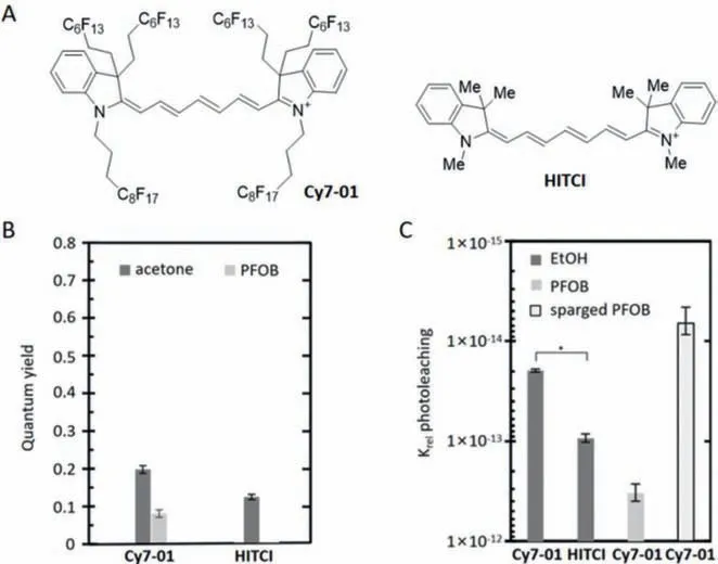
Fig.2.(A) The structure of Cy7-01 and HITCI.(B) Quantum yields of Cy7-01 and HITCI in acetone and Cy7-01 in perfluorooctyl bromide (PFOB).(C) Relative photobleaching rates of Cy7-01 and HITCI in EtOH,Cy7-01 in PFOB,and Cy7-01 in N2-sparged PFOB.*P <0.0001.Reproduced with permission [48].Copyright 2020,American Chemical Society.
Slikboeretal.[49] synthesized a new cyanine dye derived from tetridazine in the same year.This dyeCy7-02can serve as a targeted photoacoustic (PA) imaging probe for tumor imaging (Fig.3A),and it has high albumin binding properties to promote tumor localization and decomposition of cyanine dyes,enhancing its signal outputinvivo.They conductedinvivoandin vitrotumor imaging experiments to investigate the effects of this dye on tumor imaging.The fluorescence signal in the tumor was clearly visible one hour after injection ofCy7-02,and the uptake of the dye was evident in theinvitroimaging of the tumor (Fig.4A).To image calcium accumulation,they synthesized a novel PA probe,Cy7-03(Fig.3A),using bioorthogonal reactions betweenCy7-02andtrans-cyclooctene-bisphosphonate (TCO-BP).TCO-BP targets radiolabeled tetrazines to high calcium accumulation sites.In aninvivocalcium accretion imaging study,only the knees of mice withCy7-03showed strong signals (Fig.4B).Moreover,the authors used inverse electron demand Diels Alder reaction (IEDDA) to linkCy7-02to [99mTc]Tc-TCO-BP (Fig.3B),to quantitatively determine the systemic biodistribution of PA imaging dyes.

Fig.3.(A) The structures of Cy7-02 and Cy7-03.(B) The synthesis of Cy7-04.

Fig.4.(A) 143B PA images of human osteosarcoma mouse xenograft tumors injected with Cy7-02.(B) PA images of the focused knee joint 1 h after Cy7-02 injection (top)and Cy7-03 injection (bottom) are shown in vitro.Reproduced with permission [49].Copyright 2020,American Chemical Society.
According to the studies conducted,it has been demonstrated that the PA imaging probes developed by Slikboeretal.can be effortlessly imagedinvivo,and the creation of such probes is uncomplicated.Furthermore,the targeting agent can be replaced in the final stage without a complete resynthesis and optimization of the new complex.This offers a novel way of treating cancer and other ailments.
Even though the far infrared is advantageous over the near infrared,most fluorescent probes currently in existence use tetrazines that are quenched by through-bond energy transfer(TBET) or Forster resonance energy transfer (FRET) between the acceptor and donor fluorophore.However,some fluorescent probes cannot be quenched by tetrazines through energy transfer due to poor overlap with the far-infrared spectrum.In 2016,Wuetal.[50] developed a near-infrared fluorescent probe for tetrazine reactions that uses an alternative quenching mechanism.The team employed an internal charge transfer (ICT) process to quench the fluorophore,and the quenched ICT fluorophore can be revealed using a bioorthogonal click release strategy.
To determine the design strategy of probes,the authors first preparedCy7-06using the compoundCy7-05(Fig.5A).Upon addingCy7-05andCy7-06to the phosphate buffer saline (PBS)solution,the fluorescence intensity ofCy7-06was 70 times greater than that ofCy7-05.Additionally,the transfer of the absorption peak ofCy7-06from 620 nm to 560 nm after capturing it with vinyl ether confirms the logic of the ICT process.Next,the authors synthesizedCy7-07(Fig.5A) and linked it to RNA to create a probe.Finally,the team used RNA probes to label RNA in cells and imaged the cells (Fig.5B).This new mechanism can be applied to a wider range of fluorescent preparations.

Fig.5.(A) The structures of Cy7-05,Cy7-06 and Cy7-07.(B) RNA probes label cells capable of expressing green fluorescent protein.Reproduced with permission [50].Copyright 2016,American Chemical Society.
Constructing NIR probes with multiple functional components is challenging due to the instability of common NIR dyes.However,researchers are actively exploring solutions in this field.In 2020,Wangetal.[51] developed a novel NIR scaffold with three clickable handles,which exhibited favorable fluorescence properties when connected to different biomolecules.The authors utilized this scaffold to designCy7-09(Fig.6A),which was applied to A549 cells expressing cetuximab.The experimental results demonstrated thatCy7-09enabled cells with terminal alkyne groups on the cell membrane to exhibit bright NIR signals (Fig.6B),achievinginsituNIR labeling of antibodies on the surface of living cells.Additionally,the authors designed NIR-positron emission tomography (PET) dual-modal probes forinvivotumor imaging to address the challenge of introducing radiotracers with short half-lives.The authors synthesizedCy7-10(Fig.7A) and employed the probe to image U87MG tumors in naked mice,revealing a strong fluorescent signal (Fig.7B) at the tumor site.This unique scaffold with three clickable handles and continuous click reactions under biocompatibility conditions successfully resolved the issue of building NIR probes with multiple biofunctional arms.

Fig.6.(A) The structures of Cy7-08 and Cy7-09.(B) A549 cells were imaged with (top) and without (bottom) Cy7-09 labeling.Reproduced with permission [51].Copyright 2020,American Chemical Society.

Fig.7.(A) The structure of Cy7-10.(B) PET images (left) and fluorescent images (right) of U87MG tumor xenograft mice obtained at different times after intravenous injection of Cy7-10.Reproduced with permission [51].Copyright 2020,American Chemical Society.
In certain cases,some reagents cannot dissolve in water and need organic reagents at higher concentrations,otherwise their activity may decrease,or they may become more vulnerable to high temperatures.In 2016,Rodriguez [52] developed a probe that is highly water-soluble and compatible with multimode imaging.They connected the boron fluoride trap derivative to Cy7 through a bioorthogonal ligation method to obtain a multimodal imaging probe capable of [18F]-PET and near-infrared fluorescent (NIRF)imaging (Fig.8).
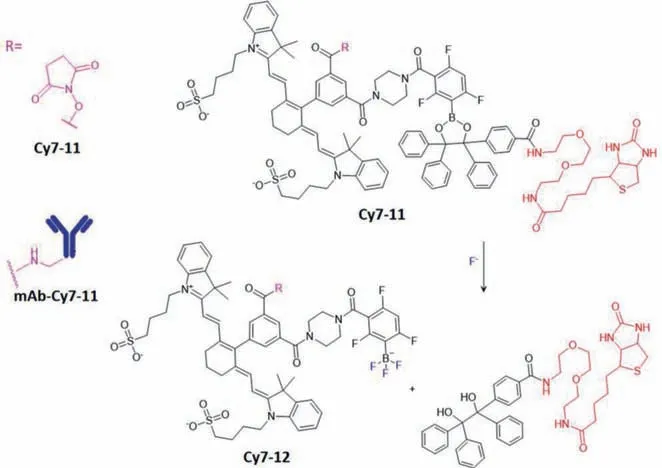
Fig.8.The structural formulas of Cy7-11 and Cy7-12 are shown in the figure.By washing with [18F]-fluorine,Cy7-11 can be decomposed into Cy7-12 and separated from the solid carrier.
Streptavidin-agarose was employed as a solid carrier for the probe after it was conjugated to the antibody.The use of fluoride aqueous solution enabled the elution of the probe,Cy7-11,from the solid phase,yielding freeCy7-12for tracking the antibody (Figs.9A and B).The residualCy7-11could be reserved for future use.The substrate could be recycled for multiple synthesis cycles by simply rinsing it with [18F]-fluoride.Additionally,the probe-antibody complex that remained uneluted by [18F]-fluoride stayed in the solid phase,ensuring the purity of the probe in the solution and increasing its specific activity.
The probe takes advantage of both solid phase tracer generation and the unique attenuation properties of [18F]-PET nuclides,resulting in a superior PET/NIRF multimodal imaging approach.This approach offers improved depth-of-penetration,spatial resolution,and temporal resolution when compared to a simple multimodal response.Additionally,the mild solid adhesion and fluorineinduced decomposition of aryl dioxyborane do not impact antigenspecific binding,allowing for the targeting of more complex biological targets.By using monoclonal antibody (mAb),this probe eliminates the need to separate multiple active species and does not require high radiochemical yields like traditional markers.Antigenbinding is unaffected by chemical attachment,solid carrier solidation,or fluorine-containing solvent elution,as demonstrated by images of [18F]-mAb-Cy7-12and [19F]-mAb-Cy7-12under controlled conditions (Fig.10).
Once the results are obtained,it is necessary to verify the binding of the antigen.In the experimental group,PC3 cells were labeled with [19F]-mAb-Cy7-12and then rinsed with unlabeled mAb,resulting in a significant reduction in membrane fluorescence,thus confirming the specificity of the [19F]-mAb-Cy7-12membrane binding to the antigen (Figs.10A and B).Although the weight of the [19F]-mAb-Cy7-12was reduced by TCEP,resulting in severe fluorescence loss,the monoclonal antibodies used in this study (Figs.10A and C) showed equivalent Cy7 fluorescence through other pathways,indicating that the binding of [19F]-mAb-Cy7-12to the antigen was necessary for endogenous fluorescence(Figs.10C and D).The experimental group incubated the cells for a longer time (Fig.10E),and performed fluorescent imaging of the cytoplasm and cell membranes with red fluorescent protein (RFP)and DiO membrane dye,respectively.The imaging results were superimposed with Fig.10E to obtain Figs.10F-I.The experimental results confirmed that increasing the incubation time could promote endocytosis.Furthermore,the experimental group demonstrated that [19F]-mAb-Cy7-12can perform fluorescence imaginginvivoat short time points and can be used to monitor tumor metastasis (Fig.9C).

Fig.9.(A) Imaging of streptavidin-agarose-solid rotating column containing biotinmAb-Cy7-11 under bright conditions and fluorescence.(B) mAb-Cy7-11 and mAb-Cy7-12 analyzed by sodium dodecyl sulfate polyacrylamide gel electrophoresis(SDS-PAGE).(C) Image of [19F]-mAb-Cy7-12 in the primary tumor and stomach at 6 h and 48 h,respectively.Reproduced with permission [52].Copyright 2016,American Chemical Society.

Fig.10.Imaging of [18F]-mAb-Cy7-12 and [19F]-mAb-Cy7-12 under a variety of controlled conditions.Reproduced with permission [52].Copyright 2016,American Chemical Society.
The fluorescence of current tetraazine-based bioorthogonal probes decreases significantly as their emission wavelength shifts to the NIR region,severely limiting their applicability in living cells and tissues.However,in 2022,Mao and colleagues [53] utilized the photoinduced electron transfer mechanism (Fig.11A) [54] to reduce background fluorescence and created a series of tetrazinebased far red/NIR fluorescent dyes with outstanding performance at longer wavelengths.One of these dyes,Cy7-13(Fig.11B),was synthesized by the authors and its fluorescence quenching was attributed to the photoinduced electron transfer mechanism.The probes produced through this technique are highly biocompatible and photostable,making them well-suited for imaging specific intracellular targets and tumorsinvivo.

Fig.11.(A) The PET process is represented by the state-crossing from a Locally Excited to an Electron Transfer State (SLEET) model.(B) The structure of Cy7-13.Note:ET for the electron transfer state;CT for the charge transfer state;LE for the locally excited state.Reproduced with permission [54].Copyright 2021,American Chemical Society.
Recently,a new strategy for designing cyanine dyes for tumor imaging was developed by Zhangetal.in 2022 [55].They utilized the torsion-induced disaggregation (TIDA) phenomenon to designCy7-14,which was then connected to tetrazine-transcyclooctene(Tz-TCO) to generateCy7-15(Fig.12A).The conformational change of the fluorophore’s heptamethyl chain from S-transto S-cisresulted in significant fluorescence enhancement,making the molecule ideal for tumor imaging.The authors successfully usedCy7-14for live-cell imaging and in a 4T1 tumor-bearing mouse model,as shown in Fig.12B and Fig.13,respectively,confirming its effectiveness for enhancing fluorescent signal and imaging tumors.

Fig.12.(A) The structures of Cy7-14 and Cy7-15.(B) Confocal laser scanning microscopy (CLSM) imaging in 4T1 cells after different concentrations of TCO treated with Cy7-14.Reproduced with permission [55].Copyright 2022,Springer Nature.

Fig.13.(A) CLSM imaging of 4T1 cells with Cy7-14 under different conditions.(B)Confocal fluorescence images of Cy7-14 and Cy7-15 in 4T1 cells.(C) NIR imaging of mouse tumors injected with RGD-TCO and Cy7-14.Reproduced with permission[55].Copyright 2022,Springer Nature.
Cy7 is frequently utilized as a fluorescent reagent in fluorescence imaging due to its exceptional biocompatibility and NIR fluorescence qualities.Fluorescence imaging can be employed for visualization in the antibody-drug conjugate (ADC) approach,utilizing photochemical properties.In 2017,Nani [56] demonstrated that the compoundCy7-16(Fig.14A) facilitated small molecule release under light irradiation within the 690 nm range.However,the compound requires improvement for clinical use.Firstly,the modification structure of the original compound should be altered to increase itsλmax,as the excitation wavelength is still relatively far from the near infrared band.Secondly,the scaffold of the initial compound must be enhanced to address the background hydrolysis effect,strengthen photooxidation,increase the effectiveness of the payload molecule,and realize high-strength labeling.

Fig.14.(A) The structures of Cy7-16,Cy7-17 and Cy7-18.(B) Fluorescence images at 800 nm.(C) Bioluminescence images of luciferase activity.Reproduced with permission[56].Copyright 2017,American Chemical Society.
After a series of explorations,the team identifiedCy7-17aandCy7-17b(Fig.14A) as potential small molecule delivery agents that required further evaluation.These compounds exhibited high redshifts and significantly reduced background hydrolysis effects.To improve efficacy and simplify conjugation processes,the team utilized bioorthogonal reactions to modify theCy7-17aandCy7-17bscaffolds and coupled them with panitumumab,a monoclonal anti-EGFR antibody commonly used in clinical settings,resulting in the final target moleculesCy7-18aandCy7-18b(Fig.14A).Cy7-18bwas found to possess a better therapeutic index and NIR photosensitivity under identical light source conditions and was therefore selected forinvivostudies.Subsequently,the team evaluated theinvivoefficacy ofCy7-18bin mice bearing MDA-MB-468-luc tumors.Through fluorescence imaging and luminescence imaging of luciferase activity (Figs.14B and C),they discovered that the drug was well-tolerated by mice and inhibited tumor proliferation.This approach provides a unique platform for cyanine scaffold remodeling to target drug delivery and offers new possibilities for treating diseases.
3.Conclusion
In conclusion,this review provides a comprehensive summary of the current applications of Cy7-type dyes in the field of bioorthogonal chemistry in terms of dye molecular structure,reaction conditions,fluorescence imaging,and bioapplications.Although Cy7-type dyes demonstrate unparalleled biocompatibility and excellent bioimaging performance in living cell,tissue andin vivo,their disadvantages in terms of photostability,fluorescence quantum yields,and toxicity remain the greatest barriers to their further application.These are still the directions we need to work on in the coming research.Furthermore,to further enhance their function and applications,some Cy7-type dyes with novel backbones will be developed and used in the future,especially those with more functionalization sites,which will provide more options for bioorthogonal chemistry.We also believe that bioorthogonal reactions with NIR fluorescence characteristics will be increasingly used in living organisms [57-60].In particular,it will play an increasingly important role in exploring the functions of various biomolecules in cellular and physiopathological processes,and clinical diagnosis and treatment.
Declaration of competing interest
The authors declare that they have no known competing financial interests or personal relationships that could have appeared to influence the work reported in this paper.
Acknowledgments
This work was supported by the National Key R&D Program of China (No.2022YFA1207400),National Natural Science Foundation of China (No.22274061),the 111 Project (No.B17019)and Fundamental Research Funds for the Central Universities (No.CCNU22QN007).
 Chinese Chemical Letters2024年2期
- Chinese Chemical Letters的其它文章
- The 3rd Xihua Chemistry and Biomedicine Forum
- Professor Hualiang Jiang: A tribute to an esteemed visionary chemist and pharmacist
- Recent advances in visible light-mediated chemical transformations of enaminones
- Development of porphyrin-based fluorescent sensors and sensor arrays for saccharide recognition
- Recent advances of versatile reagents as controllable building blocks in organic synthesis
- Synthetic host-guest pairs as novel bioorthogonal tools for pre-targeting☆