
Self-assembly 2D plasmonic nanorice film for surface-enhanced Raman spectroscopy*
2021-11-23 07:30TingtingLiu柳婷婷ChuanyuLiu劉船宇JialingShi石嘉玲LingjunZhang張玲君XiaonanSun孫曉楠andYingzhouHuang黃映洲
Chinese Physics B 2021年11期
Tingting Liu(柳婷婷), Chuanyu Liu(劉船宇), Jialing Shi(石嘉玲), Lingjun Zhang(張玲君),Xiaonan Sun(孫曉楠), and Yingzhou Huang(黃映洲)
State Key Laboratory of Coal Mine Disaster Dynamics and Control and Chongqing Key Laboratory of Soft Condensed Matter Physics and Smart Materials,College of Physics,Chongqing University,Chongqing 400044,China
Keywords: surface plasmon,surface-enhanced Raman spectroscopy(SERS),nanorice,2D plasmonic film
1. Introduction
With the fast development of nanofabrication technology in recent decades, the optical feature of metal nanostructure has attracted plenty of attention from researcher.This is mainly because the collective oscillation of free electrons in metal nanostructure could be excited by light,which is called surface plasmon.[1]This collective oscillation reaches resonance at suitable frequency of incident light,which could greatly confine light at metal surface with region at nanoscale and is also called surface plasmon resonance (SPR).[2,3]This strongly confined light at surface of noble metal nanostructure has tremendous applications, e.g.,sensing,[4]photocatalysis,[5]waveguide beyond diffraction limit,[6]and structural color,[7]etc. Among them,surface enhanced Raman spectroscopy(SERS)is one of the most interesting topics at recent years.[8-10]The huge enhanced electromagnetic field generated by surface plasmon greatly magnify the Raman intensity of the molecule adsorbed at metal surface,which overcomes the weak scattering cross section in the normal Raman.[11,12]Therefore,as a spectrum sensing technique with high sensitivity even at single molecule level,SERS has been applied in lots of fields, such as food security,[13]environmental pollution,[14]biological medicine,[12,15]criminal investigation,[16]and performance diagnosis.[17,18]
For its great influence on the enhancement and uniformity of SERS signals, the studies on SERS substrate consisted of metal nanostructure could be a core mission in SERS application. Since the status of surface plasmon is largely determined by the size, shape, and material, various metal nanostructures with different configuration have been investigated for SERS substrate, e.g., nanoparticle aggregating,[19]nanohole/pillar,[20]and nanostructure array.[21]In recent years,self-assembly 2D nanoparticle film exhibits wide application prospects in SERS substrate for its convenient fabrication process, uniformity of signals, high sensitivity and low cost.[14,22-25]Obviously, the SERS performance of 2Dplasmoninc film is dominated by the excitation and coupling of surface plasmon in nanoparticles,which is strongly influenced by the shape of nanoparticle. Therefore, the research on 2D plasmonic film consisted of novel nanoparticles as SERS substrate could be an interesting task in related fields.[13,14,26,27]
Chemical synthesized Ag nanorice is an interesting anisotropic plasmonic nanoparticle with high electromagnetic field enhancement at sharp tips, which exhibits novel surface plasmon properties such as multipolar plasmon resonances,[28,29]plasmonic circular dichroism,[30]and plasmonic Fano resonances.[31]In this work,the Ag nanorice has been adopted to self-assemble 2D plasmonic film as SERS substrate. And the SERS spectra of various molecules have been obtained on this 2D nanorice film. Interestingly,the collected spectra point out this 2D plasmonic film consisted of anisotropic nanostructures has no obvious SERS polarization dependence. Furthermore, the surface plasmon coupling has been analyzed by the simulated electric field distribution with different wavelength, which figures out this 2D nanorice film has an outstanding enhancement at near-infrared region and benefits the SERS detection of bio-sample usually excited by 785 nm laser.[8]
2. Methods and materials
All chemical reagents were purchased without further purification. The nanorice was fabricated according to Liang’s work.[32]And the 2D nanorice film was achieved by improving the interfacial strategy reported by Liuet al.,[33]as depicted in Fig.1(a). Typically,Ag nanorice colloid(5 ml)was centrifuged and washed with ethanol more than three times at 10000 rpm for 5 minutes. And then, the nanorice (0.5 ml)were redispersed in deionized water(2 ml)in a glass vial. Nhexane(1 ml)and ethyl alcohol(1 ml)were dropped into the glass vial successively to produce the organic-water interface.After induction of ethanol,a 2D Ag nanorice film was formed immediately at the organic/water interface. This 2D nanorice film was transferred onto a Si wafer for SERS measurement after N-hexane was evaporated completely(5-8 minutes).
The SERS spectra were measured by the self-built Raman spectrometer excited by 633 nm laser whose power is 0.7 mW at 100× objective. For a typical SERS measurement, 5 μL of methylene blue(MB)aqueous solution with different concentrations was dropped onto 2D nanorice film. And then the SERS spectra were performed immediately after drying it at room temperature.
3. Results and discussion
As shown in Fig.1(b),a 2D Ag nanorice film with good uniformity is obtained at the N-hexane/water interface through interfacial self-assembly method. The SEM image points out this 2D plasmonic film is arranged tightly and evenly with random orientations,which is consisted of uniform Ag nanorices(about 450 nm in length and 80 nm in width).
The SERS spectra collected on 2D nanorice film are shown in Fig. 2. The SERS spectra of MB molecule at different concentrations (10?6M, 10?7M, 10?8M, and 10?9M) exhibit apparent Raman characteristic peaks of MB at 770 cm?1, 1390 cm?1, 1430 cm?1, and 1618 cm?1, respectively. And the intensities of these Raman peaks decrease as the concentration of molecule is reduced gradually, which shows a SERS detection limit at 10?9M. The quantitative analysis of SERS intensities related to the concentrations of molecule is investigated here. The intensities of main characteristic Raman peak at 1618 cm?1as a function of MB concentrations are illustrated in Fig. 2(b). These data show a linear relationship expressed quantitatively asI=8688.27logC+73850,in whichCis the concentration of MB and I is the SERS intensities at 1618 cm?1.

Fig.1. (a)The fabrication process of 2D Ag nanorice film. (b)The optical image of obtained 2D Ag nanorice film and its corresponding SEM image.(c)SEM image of individual Ag nanorice.
The linear regression (R2) of this expression is equal to 0.961, which indicates this curve has a high degree of linear fitting. This result points out the quantitative detection of analyte could be performed on this 2D nanorice film. The SERS uniformity of this 2D nanorice film is also studied in this work.SERS spectra of MB molecule are collected at 36 positions on 2D nanorice film in two perpendicular direction with 5μm interval,whose intensities at 1618 cm?1Raman peak are shown in Fig. 2(c). Obviously, the Raman signals are enhanced at all positions in this 30 μm×30 μm mapping region, whose relative standard deviation (RSD) is less than 10%. This experiment data demonstrate our 2D nanorice film exhibits good uniformity of SERS enhancement. Furthermore,different analytes are also studied on this SERS substrate,whose data are shown in Fig. 2(d). The SERS spectrum of R6G at 10?7M(violet line) exhibits its own characteristic Raman peaks at 1306 cm?1, 1353 cm?1, 1504 cm?1, and 1565 cm?1, respectively. And the SERS spectrum of p-aminothiophenol(PATP)10?7M(orange line)shows interesting Raman peaks at 1386 cm?1, 1428 cm?1, and 1577 cm?1, which belong toρ,ρ'-dimercaptoazobenzene(DMAB)molecule.This demonstrates surface plasmon driven catalysis reaction occurs,where the PATP is converted into DMAB and similar to the previous reports.[34]SERS detection of analyte in gas phase is also executed on this 2D nanorice substrate. The analyte is formaldehyde (CH2O), a common harmful gas in human life, which is often diffused from new furniture. To simulate the diffusion of formaldehyde gas,the corresponding experiment setup is shown in Fig.S1 in the supporting materials based on Yang’s work.[19]Firstly,both the formaldehyde solution heated at 35°and 2D nanorice film are located in a chamber for 10 minutes.And then,the film is performed SERS measurement immediately without any other experiment procedure. In Fig. 2(d),the obvious SERS peaks(blue line)at 910 cm?1,1081 cm?1,1325 cm?1and 1484 cm?1are consistent with the characteristic Raman peaks of formaldehyde solution,[35]which point out the diffused formaldehyde gas in chamber is detected by our 2D nanorice film. This result demonstrate the application prospect of our film as SERS substrate for gas detection.
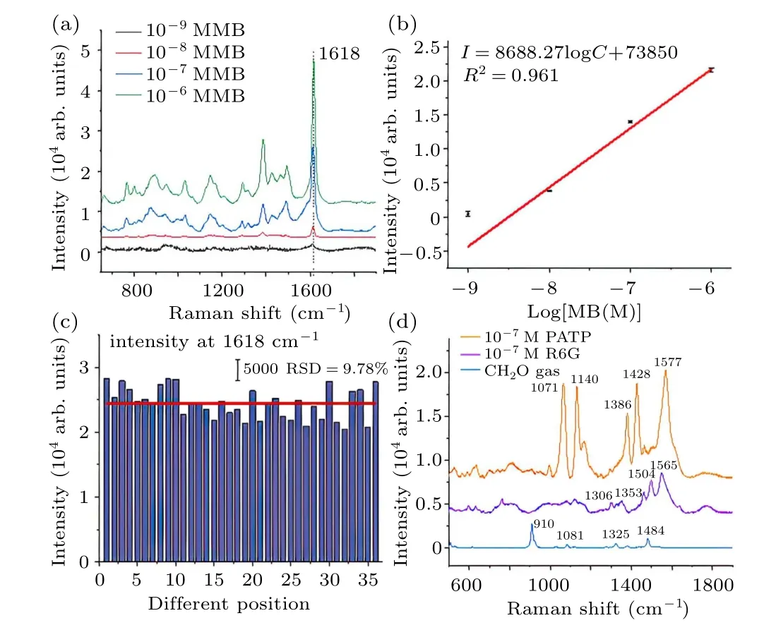
Fig. 2. (a) The SERS spectra of MB molecules at various concentrations. (b)SERS intensity at 1618 cm?1 as a function of MB concentrations.(c)SERS intensitis at 1618 cm?1 of MB collected at various positions on 2D nanorice film. (d)The SERS spectra of R6G,PATP and formaldehyde gas.
Since nanorice is a classic anisotropic structure, the polarization dependence of this 2D nanorice film as SERS substrate is studied. The reflection spectra with different polarizations of incident light is illustrated in Fig. 3(a). Interestingly,there is no obvious polarization dependence of incident light shown in reflection spectra of this 2D nanorice film. The reason could be that the anisotropic property of nanorice is eliminated by the random orientation of nanorice in 2D film as shown in SEM image in Fig. 1(b). Apparently, there is an obvious SPR peak at about 780 nm in reflection spectrum of 2D nanorice film,which is quite different from the SPR peak exhibited in extinction spectrum of nanorice colloid shown in Fig. S2 in the supporting materials. This difference could be understood by the complicated surface plasmon coupling between the adjacent Ag nanorices in 2D film while it seems as individual nanoparticle in nanorice colloid. The SERS spectra of MB collected on this 2D nanorice film with various polarization of incident light is shown in Fig. 3(b). It is clear that there is no variation in SERS intensities with different polarization. The detail experiment data are shown in Fig.3(c)that the intensities of characteristic Raman peak at 1618 cm?1are plotted as a function of the polarization of incident laser in polar coordinates. The obvious circle (blue line) is fitted by the experiment data (red dots) whose RSD is about 7.28%.The data above figure out our 2D plasmonic film consisted of anisotropic nanorice has no obvious polarization dependence,which benefits its application in SERS measurements.
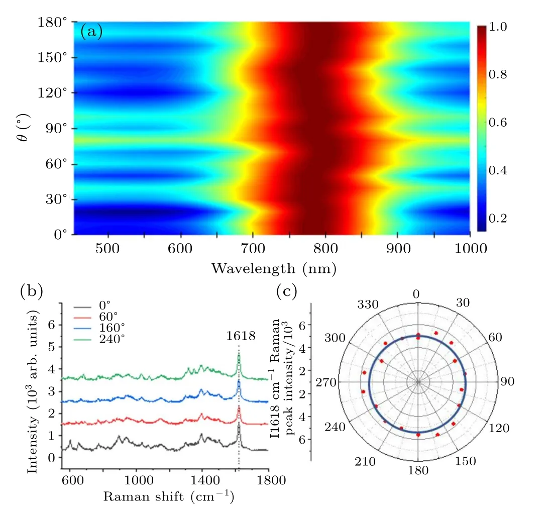
Fig. 3. (a) The reflection spectra of 2D Ag nanorice film with different polarizations. (b)The SERS spectra of MB molecules with different polarizations. (c)The SERS intensities at 1618 cm?1 with various polarizations.
To further understand the surface plasmon properties of this 2D nanorice film, the electric field distribution is investigated through Comsol multiphysics based on the finite element method. Since configurations of nanorices in 2D film are complicated as shown in Fig. 1(b), the simulation model is simplified to nanorice dimer to study the surface plasmon coupling between nanoparticles. Because all SERS spectra excited by 633 nm laser and SPR wavelength in reflection spectrum is about 780 nm, the electric field distributions of nanorice dimer at 633 nm and 785 nm (wavelength of common Raman laser)are shown in Fig.4,where the light propagates along thezdirection and its polarization is parallel to thexdirection. There are two configurations of nanorice dimer(450 nm in length and 80 nm in width) and found that configuration A is middle to middle while configuration B is tip to tip. As shown in Figs.4(a)-4(d),obvious electric field enhancement is obtained in nanorice dimer gap. This enhancement comes from the surface plasmon coupling between two nanorice,which is confirmed by the simulated surface charge distribution in Fig. S3 in the supporting materials. The situation with polarization parallel to -y-direction is shown in Fig.S4 in the supporting materials,in which there is no obvious enhancement for weak surface plasmon coupling between nanoparticles.
Apparently, the electric enhancement at 785 nm in nanorice dimer with both two configurations is much greater than that at 633 nm, where the maximal enhancement is 9.17 and 19.8 at 633 nm while is 40.3 and 187 at 785 nm, respectively. This result could be understood by the SPR peak at about 780 nm shown in reflection spectrum that much larger enhancement could be generated at resonance condition.Since the SERS intensity is in direct proportion to biquadratic electric field enhancement,an interesting finding is obtained based on the simulation results that the SERS signals is dominated by molecules adsorbed at different regions of nanorice surface at various wavelengths. Specifically, the SERS intensity mainly comes from molecules adsorbed at middle of nanorice(~57%) excited by 633 nm laser and molecules adsorbed at tip (~97%) excited by 785 nm laser, whose data in detail could be seen in Fig.S5 in the supporting materials.

Fig.4.Electric field distributions of nanoparticle dimer,nanorice dimer with configuration A(a)at 633 nm,(b)at 785 nm,nanorice dimer with configuration B(c)at 633 nm,(d)at 785 nm,nanosphere dimer,(e)at 633 nm,(f)at 785 nm.
To verify the influence of nanoparticle shape on the SERS enhancement of 2D plasmonic film, electric field distribution of Ag nanosphere dimer is also simulated and shown in Figs. 4(e)-4(f). The diameter of nanophere is 80 nm which is equal to the width of nanorice with other simulated conditions remained the same. Apparently, the enhancement is generated in gap that it is 83.5 at 633 nm and 50 at 785 nm.Contrast to nanorice dimer, the enhancement of nanospeher dimer is much greater at 633 nm. Furthermore, the maximal enhancement of nanosphere dimer at 785 nm is much weaker than that of nanorice dimer,which is 50/40.3 in configuration A and 50/187 in configuration B.Considering the SERS intensity is in direct proportion to biquadratic electric field enhancement and all configurations obtained in our film, the conclusion could be obtained that the 2D Ag nanorice film exhibits a good application prospect in near IR SERS,which benefits the SERS monitoring in biological field usually excited by 785 nm laser.
4. Conclusion
In this work, self-assembly 2D plasmonic film consisted of Ag nanorice is fabricated and its SERS performance is investigated. The collected SERS spectra of various molecule on this 2D nanorice film demonstrate quantitative detection could be performed on this SERS substrate without polarization dependence of incident laser. The simulated electric field and surface charge density distribution point out the SERS enhancement of this 2D nanorice film comes from the surface plasmon coupling between nanorices and the SERS signals is dominated by molecules adsorbed at different regions of nanorice surface at various wavelengths,e.g.,~97%SERS signal come from molecules adsorbed at tips excited by 785 nm laser. Compared to common 2D nanosphere film,the data figure out our 2D nanorice film could be a good near IR SERS substrate, which benefits the bioanalysis with 785 nm laser.
- Chinese Physics B的其它文章
- Numerical investigation on threading dislocation bending with InAs/GaAs quantum dots*
- Connes distance of 2D harmonic oscillators in quantum phase space*
- Effect of external electric field on the terahertz transmission characteristics of electrolyte solutions*
- Classical-field description of Bose-Einstein condensation of parallel light in a nonlinear optical cavity*
- Dense coding capacity in correlated noisy channels with weak measurement*
- Probability density and oscillating period of magnetopolaron in parabolic quantum dot in the presence of Rashba effect and temperature*