
Study on the mechanism of herb cake-partitioned moxibustion inhibiting tumor growth in colitis-associated colorectal cancer based on KDM4D receptor
2024-02-27 08:24XUHongxiao徐宏瀟WANGYiran王逸然XUJing徐靜HUANGYan黃艷QIAOYu喬宇LIFeifei李斐斐LIGuona李國娜ZHAOJimen趙繼夢LIFeng李峰CHENZiyi陳子怡LIKunshan李昆珊GUMuen顧沐恩WUHuangan吳煥淦
XU Hongxiao (徐宏瀟), WANG Yiran (王逸然), XU Jing (徐靜), HUANG Yan (黃艷),, QIAO Yu (喬宇),,LI Feifei (李斐斐),, LI Guona (李國娜), ZHAO Jimen (趙繼夢),, LI Feng (李峰), CHEN Ziyi (陳子怡),,LI Kunshan (李昆珊),, GU Mu’en (顧沐恩),, WU Huangan (吳煥淦),
1 Yueyang Hospital of Integrated Traditional Chinese and Western Medicine, Shanghai University of Traditional Chinese Medicine, Shanghai 200437, China
2 Shanghai Research Institute of Acupuncture and Meridian, Shanghai 200030, China
Abstract Objective: To explore the inhibitory mechanism of herb cake-partitioned moxibustion on tumor growth in colitisassociated colorectal cancer (CAC) based on histone lysine demethylase 4D (KDM4D).
Keywords: Point, Qihai (CV6); Point, Tianshu (ST25); Medicinal Cake-partitioned Moxibustion; Colitis-associated Neoplasms; Colorectal Neoplasms; KDM4D; Wnt/β-catenin Signaling Pathway; Rats
Colitis-associated colorectal cancer (CAC) is closely related to recurrent colitis caused by ulcerative colitis(UC) and Crohn disease (CD)[1].Both UC and CD belong to inflammatory bowel diseases (IBD).The canceration caused by UC accounts for 1%-2% of all colorectal cancer, which is higher than the carcinogenic rate of CD.Moreover, CAC patients caused by UC are younger, with more descending colon involvement and multifocality[2].Moreover, 10%-15% of IBD patients died of CAC[3].At present, the risk factors of IBD developing into CAC include onset age, course of disease, mucosal inflammation degree, lesion scope, and related family history.UC patients with a long course of disease are at greater risk of CAC.With the prolonged disease course,the cumulative risk of UC-related CAC is as high as 18%-20%[4].
At present, it is believed that the pathogenesis of CAC mainly involves chronic inflammation of colorectal mucosa, which causes intestinal mucosal barrier destruction to increase the damage to intestinal epithelial cells and tissues.The repeated local damage turns into atypical hyperplasia and finally develops into tumors[5].Studies have shown that CAC is less, and the expression levels of C-myc oncogene, cyclin D1 (phopho Thr286), proliferating cell nuclear antigen (PCNA), and β-catenin are down-regulated under the knockout condition of histone lysine demethylase 4D (KDM4D)gene versus the wild type.KDM4D inhibitor inhibits colonic tumor growth in wild-type CAC mice.KDM4D is a key protein that promotes the activation of Wnt/βcatenin signaling pathway in CAC[6].Abnormal activation of the Wnt/β-catenin pathway by the adenomatous polyposis coli (APC) gene and the somatic axis inhibitor(Axin)[7]regulates the expression levels of key factors,such as KDM4D, Axin, and APC, to inhibit the Wnt/βcatenin pathway activation in CAC, which is of great significance in delaying tumor growth.
Moxibustion is a characteristic treatment of traditional Chinese medicine.Our group previously found that herb cake-partitioned moxibustion and ginger-partitioned moxibustion effectively treated IBD and reduced colitis[8-10]; moxibustion inhibited the growth of CAC tumors through Wnt/β-catenin signaling pathway[11-13].In this study, the KDM4D expression, cell proliferation, apoptosis-related proteins, and related upstream (APC and Axin) and downstream molecules of Wnt/β-catenin pathway were observed in CAC rat model with KDM4D inhibitor as the control.The mechanism of herb cake-partitioned moxibustion inhibiting colonic tumor growth in CAC rats was discussed based on the KDM4D and Wnt/β-catenin signaling pathways.
1 Materials and Methods
1.1 Laboratory animal
Male Sprague-Dawley rats with a body mass of(100±20) g were raised in the Experimental Animal Center of Yueyang Hospital of Integrated Traditional Chinese and Western Medicine, Shanghai University of Traditional Chinese Medicine [License No.SCXK (Hu)2017-0005].The feeding environment was 12 h/12 h circadian rhythm, with a room temperature of (20±2) ℃and an indoor humidity of 50%-70%.The rats were fed adaptively for one week before experiments.All experimental operations strictly followed theGuiding Principles for the Care of Experimental Animals.This study was approved by the Ethics Committee of Yueyang Hospital of Integrated Traditional Chinese and Western Medicine, Shanghai University of Traditional Chinese Medicine (Ethic No.YYLAC-2021-117).
1.2 Main reagents and instruments
Antibodies against rat PCNA and APC (Cat.No.ab29,No.ab40778, Abcam, China); rat cleaved caspase3 antibody (Cat.No.9661T, CST, USA); antibodies against rat Axin1, JMJD2D, and Trizol reagent (Cat.No.MA5-14854, No.PA5-100762, No.15596018, Thermo Fisher Scientific Inc., USA); horseradish peroxidase labeled goat anti-rabbit immunoglobulin G (Cat.No.A0208,Beyotime Biotechnology, China); azoxymethane (AOM)and dextran sulfate sodium (DSS) (Cat.No.02180139,No.00081551, MP Biomedicals, China); pentobarbital sodium (Cat.No.P3761, Sigma, USA); hematoxylin and eosin (Cat.No.D006, No.D007, Nanjing Jiancheng Bioengineering Institute, China); DEPC H2O and agarose(Cat.No.SD01005, No.SD0241, Shanghai Siding Biotechnology Co., Ltd., China); RT reagent kit and TB Green quantitative polymerase chain reaction kit (Cat.No.RR047A, No.RR420A, TAKARA, China).
ME54E/02 microelectronic balance (Mettler Toledo,China); HI1220 slide warmers and HI1210 water bath(Leica, Germany); pipette (Eppendorf, Germany); BPG-9070A electric thermostat (Shanghai Yiheng Scientific Instrument Co., Ltd., China); 5810R centrifuge (Thermo Fisher Scientific Inc., USA); F50 microplate reader (Tecan,China).
1.3 Modeling preparation and identification
Twenty-eight rats were randomly divided into a normal group (n=8) and a group for CAC model preparation (n=20).The CAC rat model was established by the combined administration of AOM and DSS.The model rats were first injected with AOM[10 mg/(kg·bw)].After 7 d, three cycles of DSS(3%-2%-2%) administration were offered via free drinking water to stimulate inflammation.The specific implementation plan was as follows: each cycle was sustained for 21 d with free diluted DSS drink for the first 4 d and then regular water for the next 17 d.DSS dilution ratios were 3%, 2%, and 2%, respectively, in the first 4 d of the three DSS cycles (Figure 1)[14].
The general condition of rats was observed and recorded in the first 4 d of each DSS cycle.After modeling, 2 rats in the normal group and 2 rats in the group for CAC model preparation were selected for model identification.Macroscopic observation showed tumors in the inner wall of colon, and tumor tissue formation on the surface of colon; dysplasia of mucosal glands and “back-to-back” phenomenon were observed under the microscope in the group for CAC model preparation, which indicated that the CAC model with adenocarcinoma formation was successfully established(Figure 2).
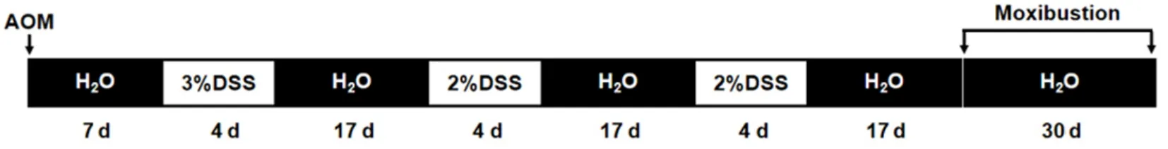
Figure 1 CAC rat model preparation

Figure 2 Identification of the CAC rat model (hematoxylin-eosin staining, ×40)
1.4 Groups
After the model was identified as a success, 6 rats in the normal group were still used as the normal group.Eighteen CAC-modeled rats were randomly divided into a CAC group, a herb cake-partitioned moxibustion group, and an inhibitor group (n=6).
1.5 Intervention methods
Rats in the CAC group and the normal group received the same fixation as those in the herb cake-partitioned moxibustion group without other interventions.
Rats in the herb cake-partitioned moxibustion group and the inhibitor group began to receive the corresponding intervention on the second day after modeling.
Herb cake-partitioned moxibustion group: Qihai (CV6)and bilateral Tianshu (ST25) were selected.Rat point positioning consulted theExperimental Acupuncturology[15].GroundFu Zi(Radix Aconiti Lateralis Praeparata) and other Chinese herbs into powder, added yellow wine and mixed, and then made a herb cake with a diameter of about 0.8 cm and a thickness of about 0.4 cm using a special mold.Moxa cones were made from moxa wool, each weighing about 90 mg.The herb cake was first placed on the point, the moxa cone was placed on the herb cake and then ignited to apply moxibustion.Two cones were used at each point, once a day for 30 d with moxibustion conducted 26 times and one day rest every week.
Inhibitor group: Rats in the inhibitor group were given the intraperitoneal injection of KDM4D inhibitor [5-chloro-8-hydroxyquinoline (5-c-8HQ)][10 mg/(kg·bw)][16], once a day for 30 d.
1.6 Animal specimen collection
The rats were anesthetized by intraperitoneal injection of 2% sodium pentobarbital solution[40-50 mg/(kg·bw)] before sampling and after fasting for 24 h.The rats were fixed on the anatomical plate in a supine position after being successfully anesthetized.The colon tissue was separated down to the anus with the cecum as the main mark, the complete colon length was measured and recorded after the abdominal cavity was opened.The colon was cut longitudinally along the midline and washed with normal saline.Observed the surface of colon tissues.Measured and recorded the diameter and quantity of colonic tumors.Three sections with a length of 1 cm were collected from the middle and lower part of the colon, and the distal part of the colon with tumors (the corresponding part of colon if there was no tumor) was fixed in 4% paraformaldehyde solution.The other two colon sections were cut into pieces, put into a cryopreservation tube, and stored in the refrigerator at -80 ℃ for later use.
1.7 Observation indicators and methods
1.7.1 General condition and tumor formation
The general conditions of rats were recorded daily,including mobility, coat color, stool pattern, and macroscopically visible bloody stool.In addition to the diameter and number of colonic tumors, the maximum diameter (a) and the minimum diameter (b) of each tumor larger than 1 mm in diameter were measured,and the tumor volume was calculated by the formula of V = (π/8) × a × b2[17].
1.7.2 Pathological observation of rat colon tissues
The distal tumor-bearing colon tissues were subjected to dehydration, embedding, continuous slicing at 4 μm, spreading, and baking.Tumor sections were dewaxed and stained with hematoxylin-eosin (HE)as follows: treating with hematoxylin staining solution for 2 min, running water washing for 10 min, 1%ethanol hydrochloride differentiation for about 5 s,running water washing for 5 min, and treating with eosin staining solution for 2 min.After that, dehydration,transparency, mounting, and observation under an optical microscope were carried out.
1.7.3 Expression levels of PCNA, cleaved caspase3,KDM4D, APC, and Axin in colon tissues
Immunohistochemistry was used to detect the protein expression levels of PCNA, cleaved caspase3,KDM4D, APC, and Axin in distal tumor-bearing colon tissues.Paraffin sections of rat colon tissues were dewaxed, hydrated, and heat-antigen repaired.Then,the autologous antibodies were inactivated and blocked,respectively.After incubation with proper diluted rabbit anti-rat primary antibodies (1:6 000 dilution for PCNA,1:400 dilution for cleaved caspase3, 1:200 dilution for KDM4D, 1:1 000 dilution for APC, 1:50 dilution for Axin)and the corresponding sheep anti-rabbit secondary antibody (dilution ratio 1:200), PBS washing was performed, followed by DAB color development,hematoxylin staining, dehydration, and mounting.
Sampling and analysis of image information: 3-5 visual fields were randomly selected under the optical microscope to collect images, and the average optical density (AOD) of images was analyzed by Image-Plus Pro 6.0 software.The AOD of each slice was calculated by AOD = Integrated optical density (IOD) ÷ Positive area of positive target (Area).
1.7.4 The mRNA expression levels of APC, Axin, cyclin D1, MMP-7, and MMP-9 in colon tissues
The mRNA expression levels of APC, Axin, cyclin D1,MMP-7, and MMP-9 were detected by real-time quantitative polymerase chain reaction (RT-qPCR).Tumor-bearing colon tissues were collected and the total RNA was extracted using the Trizol method.PCR was carried out by adding appropriate reagents to the extracted total RNA.The sequences of amplification primers are shown in Table 1.The reaction condition was 95 ℃ for 2 min, 1 cycle; 94 ℃ for 10 s, 60 ℃ for 10 s, 72 ℃ for 40 s, 40 cycles.The data were analyzed by 2-△△Ctmethod.

Table 1 Primer sequence
1.8 Statistical methods
The SPSS version 21.0 software was used for statistical analysis.All data were measurement data.Normality and homogeneity of variance were tested.Data with normal distribution and homogeneous variance were expressed as mean ± standard deviation(±s), and one-way analysis of variance was used for inter-group comparisons.The least significant difference method was used for further comparison if there were significant differences.Inter-group analysis was performed using Dunnett’s T3 for data not meeting the homogeneity of variance.The median (lower quartile,upper quartile) [M (QL, QU)] was used to describe the central trend of the data, and the nonparametric test method was used for inter-group comparisons for data not conforming to normal distribution.The size of test was set as α=0.05.P<0.05 indicated that the difference was statistically significant.
2 Results
2.1 Observation of the general condition of rats in each group
During the experiment, rats in the normal group were agile and stable with shiny fur, normal drinking and eating, ruddy around the anus, and granular stool.Compared with the normal group, rats in the CAC group were slow to move and easy to get together with dull and yellow fur, a thin body, and slow body mass gain;also a downward trend of body mass, decreased diet and drinking, blood around the anus, and thin and soft bloody stool in some rats; fishy bedding in cages and blood stains on cage walls were also found.Compared with the CAC group, rats in the herb cake-partitioned moxibustion group and the inhibitor group showed increased activities, shiny fur, rapid body mass gain,increased water drinking and diet, and gradually formed stool, while some rats still had bloody stool.
2.2 Colon length and tumor formation of rats in each group
As Figure 3A shows, the colon length in the CAC group was significantly shorter than that in the normal group (P<0.01).Compared with the CAC group, the colon length in the herb cake-partitioned moxibustion group and the inhibitor group increased significantly(P<0.01), but there was no significant difference between the latter two groups (P>0.05).
As Figure 3B demonstrates, the average number of colonic tumors in the CAC group was significantly larger than that in the normal group (P<0.01).Compared with the CAC group, the average number of colonic tumor formation in the herb cake-partitioned moxibustion group and the inhibitor group was decreased (P<0.05),but there was no significant difference between these two groups (P>0.05).
According to Figure 3C, the volume of colonic tumors in the CAC group was significantly larger than that in the normal group (P<0.01).Compared with the CAC group,the volume of colonic tumors in the herb cakepartitioned moxibustion group and the inhibitor group was reduced (P<0.05), but there was no significant difference between these two groups (P>0.05).
As Figure 3D displays, the CAC group, the herb cakepartitioned moxibustion group, and the inhibitor group showed different degrees of tumor formation with macroscopic observation, and most of the tumors were in the middle and lower part of the colon.
2.3 Pathological observation of rat colonic tumor tissues among groups
After HE staining, the histopathological features of tumor-bearing colon in each group were observed under the microscope.The colonic mucosa structure was clear, the epithelium tissue was intact, and the glands were arranged regularly and orderly without congestion, edema, or inflammatory cell infiltration in the normal group.In the CAC group, the structure of colonic mucosa was damaged; the epithelial cells were disordered with abnormal morphology, enlarged and obvious deep staining nucleus, a high proportion of nucleoplasm, and increased mitosis; there were various levels of dysplasia accompanied by basal infiltration,interstitial congestion and edema, disordered glandular duct structure, “back-to-back” and cowall phenomena,many eosinophilic masses, and exfoliated tumor cells and inflammatory infiltrating cells in the glandular cavity,indicating that the adenocarcinoma developed.Compared with the CAC group, improvements were found in the colonic epithelial cells arrangement, nuclei morphology, inflammatory cell infiltration and mucosal edema, and dysplasia in the herb cake-partitioned moxibustion group.Compared with the normal group,more colonic epithelial cells were proliferated with increased and deeply stained nuclei in the inhibitor group.The degrees of inflammatory cell infiltration and gland atrophy and dysplasia were lower in the inhibitor group than in the CAC group (Figure 4).
2.4 Tumor proliferation and apoptosis in CAC rats
The PCNA protein expression level was significantly higher, and the cleaved caspase3 protein expression level was significantly lower in the tumor-bearing colon tissues in the CAC group than in the normal group(P<0.01).Compared with the CAC group, the protein expression level of PCNA (a marker of tumor proliferation) was significantly reduced, and cleaved caspase3 (a marker of apoptosis) was significantly increased in colon tissues of the herb cake-partitioned moxibustion group and the inhibitor group (P<0.01).However, there was no significant difference between the latter two groups (P>0.05).See Figure 5 and Figure 6.

Figure 3 Comparison of colon length and tumor formation in rats among groups

Figure 4 Histopathological observation of colonic tumors (hematoxylin-eosin staining, ×200)

Figure 5 Protein expression of PCNA and cleaved caspase3 in colon tissues in each group (immunohistochemistry, ×200)

Figure 6 Comparison of PCNA and cleaved caspase3 protein expression in colon tissues among groups
2.5 Expression levels of KDM4D and Wnt/β-catenin signaling pathway proteins
Compared with the normal group, the KDM4D protein expression level in the tumor-bearing colon tissues of CAC group increased significantly (P<0.01),while the protein expression levels of APC and Axin were significantly reduced (P<0.01); compared with the CAC group, the protein expression level of KDM4D was significantly reduced (P<0.01), while the protein expression levels of APC and Axin increased significantly(P<0.01).There was no significant difference in the expression of the above proteins between the herb cake-partitioned moxibustion group and the inhibitor group (P>0.05).See Figure 7 and Figure 8.
It can be seen from Figure 9 that the cyclin D1 mRNA expression level in the tumor-bearing colon tissues was significantly higher (P<0.01), while the mRNA expression levels of APC and Axin were significantly lower (P<0.01) in the CAC group than in the normal group.Compared with the CAC group, the cyclin D1 mRNA expression level was significantly reduced(P<0.01), while the mRNA expression levels of APC and Axin were increased (P<0.05) in the herb cakepartitioned moxibustion group and the inhibitor group.The difference between the herb cake-partitioned moxibustion group and the inhibitor group was not statistically significant (P>0.05).

Figure 7 Protein expression levels of KDM4D, APC, and Axin in colon tissues of each group (immunohistochemistry, × 200)

Figure 8 Comparison of KDM4D, APC, and Axin protein expression in colon tissues among groups

Figure 9 Relative expression levels of cyclin D1, APC, and Axin mRNAs in colon tissues of each group
2.6 Regulatory effect of herb cake-partitioned moxibustion on KDM4D-mediated migration and invasion-related factors in CAC rats
As Figure 10 shows, the mRNA expression levels of MMP-7 and MMP-9 in colon tissues of the CAC group were significantly higher than those of the normal group (P<0.01).Compared with the CAC group, the mRNA expression levels of MMP-7 and MMP-9 in colon tissues of the herb cake-partitioned moxibustion group and the inhibitor group were reduced (P<0.01 orP<0.05).There was no significant difference between the herb cake-partitioned moxibustion group and the inhibitor group (P>0.05).

Figure 10 Relative expression levels of KDM4D-mediated migration and invasion related factors: MMP-7 and MMP-9
3 Discussion
CAC belongs to the categories of “intestinal slip”,“intestinal wind”, “Zang-organ poison”, “diarrhea”,“abdominal masses”, and “hematochezia” in traditional Chinese medicine[18], with abdominal pain, diarrhea,and hematochezia as the main symptoms.Moxibustion therapy is simple, economical, and practical, and has been widely used in clinical practice.In recent years, its application in colorectal cancer treatment has been increasing[19-20].The pathogenesis of CAC is mostly the deficiency of healthy Qi and Zang-Fu organs.The damp toxin accumulates in the intestine for a long time and becomes cancer with external evil attacking or improper diet.Therefore, Chinese medicine believes that the main idea to control the occurrence and development of CAC is to strengthen body resistance and eliminate pathogenic factors[21].In this study, herb cake-partitioned moxibustion was applied to Tianshu(ST25) and Qihai (CV6) to treat CAC in rats.Tianshu(ST25) is the front-Mu point of the large intestine.Its pivotal role is reflected in regulating the ascending and descending of spleen-stomach Qi, the transportation of intestine, and governing free flow of Qi of the livergallbladder.It is closely related to the movement of Qi[22].Qihai (CV6) is the place where the innate Yuanprimordial Qi gathers.It can tonify the Yuan-primordial Qi, regulate Qi movement, and have a good curative effect on internal medicine diseases, such as spleen and stomach disorders[23].The compatibility of Tianshu(ST25) and Qihai (CV6) points has the effects of inspiring lower Jiao (energizer) Yang Qi, raising the clear and lowering the turbid, invigorating the spleen, and harmonizing the stomach, tonifying the deficient, and cultivating Yuan-primordial Qi.
Previously, our research group observed the effects of herb cake-partitioned moxibustion and gingerpartitioned moxibustion on CAC from the perspectives of purine receptor P2X7, Wnt/β-catenin signaling pathway, and P2X7/STAT3/VEGF, and found that both moxibustion methods increased the protein expression level of purine receptor P2X7, inhibited the transcription of oncogene C-myc, and improved the tumor proliferation in CAC rats[12].The mechanism of moxibustion on CAC was also discussed from the perspective of cell proliferation.It was found that moxibustion could delay tumor growth and reduce the protein expression levels of PCNA and cyclin D1 in colon tissues, which preliminarily explained the mechanism of moxibustion in inhibiting CAC tumor growth[11-13].The above research results provided reliable evidence for moxibustion to prevent and treat CAC, and explained its regulation mechanism from many aspects, indicating that moxibustion has good application value in treating CAC.
Cell proliferation and apoptosis is a complex and multi-factor process.PCNA protein is involved in cell cycle regulation and plays an important role in DNA metabolism[24].The increased PCNA protein expression has a high correlation with tumor invasion, and its overexpression is related to the malignant progression of CAC and other cancers[25].Cleaved caspase3 is a cysteine protease involved in the execution stage of apoptosis.Low expression of cleaved caspase3 indicates the invasiveness and poor prognosis of cancers.On the contrary, high expression indicates that the tumor has lower invasion and development with better prognosis[26].Based on the expression levels of PCNA and cleaved caspase3, the effect of herb cakepartitioned moxibustion on the proliferation and apoptosis of tumor cells was judged here.The results showed that the expression level of PCNA protein in the CAC group was higher than that in the normal group.Compared with the CAC group, the PCNA protein expression level in the herb cake-partitioned moxibustion group and the inhibitor group was significantly reduced, while the expression level of cleaved caspase3 in colon tissues of the CAC group was significantly lower than that of the normal group.The expression level of cleaved caspase3 in the two treatment groups was significantly higher than that in the CAC group.The results suggested that both moxibustion and the KDM4D inhibitor could improve the proliferation of colonic tumors.
The occurrence and development of CAC involves multiple genes and signaling pathways, among which abnormal activation of Wnt/β-catenin signaling pathway,a key factor, plays a great role in driving the growth,invasion, and development of colon cancer cells[27],which can lead to the occurrence, development, and metastasis of CAC.APC inactivation mutations have been found in about 80% of human colonic tumors[28].APC, Axin, and GSK-3β bind to β-catenin in the cytoplasm to form polyprotein degradation complexes.This complex promotes the phosphorylation and degradation of β-catenin protein, thus maintaining the level of β-catenin at a low level and failing to start the transcription of downstream target genes[29].This negatively regulates the Wnt/β-catenin signaling pathway.When APC, Axin, and GSK-3β are inactivated,downstream genes such as cyclin D1, MMP-7, and MMP-9 are transcribed, which promotes tumor cell proliferation and inhibits apoptosis[30].Cyclin D1 promotes cell proliferation and belongs to protooncogene.Overexpression and amplification of cyclin D1 gene have been found in many tumors.There is an obvious correlation between the increased MMP family activity and the invasion and migration of tumor cells.MMP-7 and MMP-9 degrade extracellular matrix, such as basement membrane, which makes it difficult for cells to resist the invasion and migration, leading to an increase in tumor metastasis rate[31].Therefore, this study observed the activation of upstream and downstream molecules of Wnt/β-catenin signaling pathway to judge the mechanism of Wnt/β-catenin signaling pathway in CAC progression.
KDM4D is similar to KDM4A and KDM4C in function,and it promotes the proliferation and migration of colon cancer cells[32].KDM4D also acts as an antagonist of tumor suppressor gene p53 to activate Wnt/β-catenin signaling pathway, thus leading to the formation and development of liver cancer[33-34].Studies have shown that the expression levels of KDM4D and PCNA are positively correlated with the activation of Wnt/β-catenin signaling pathway in AOM/DSS-induced colorectal cancer mice, and KDM4D-knockout prevents the occurrence of AOM/DSS-induced colorectal cancer in mice[35].Therefore, it can be inferred that KDM4D is involved in the regulation of tumorigenesis.Our results showed that the herb cake-partitioned moxibustion inhibited the expression of PCNA and up-regulated the expression of cleaved caspase3, suggesting that herb cake-partitioned moxibustion might inhibit the proliferation of CAC cancer cells and promote the apoptosis of tumor cells.Previous studies of our research group have shown that herb cake-partitioned moxibustion can inhibit tumor growth by regulating the Wnt/β-catenin signaling pathway in CAC colon tissues.On this basis, we also found that herb cake-partitioned moxibustion activated the protein expression levels of APC and Axin, the upstream of Wnt/β-catenin signaling pathway, and inhibited the mRNA expression levels of cancer-promoting factor cyclin D1 and cancer invasionrelated genes (MMP-7 and MMP-9), the downstream of Wnt/β-catenin signaling pathway.Our results indicated that herb cake-partitioned moxibustion may delay tumor progression in CAC rats by inhibiting abnormal activation of Wnt/β-catenin signaling pathway.By using the KDM4D inhibitor as a control, we confirmed that herb cake-partitioned moxibustion inhibited the KDM4D expression in colon tissues of CAC rats with a similar inhibition effect on tumor growth as that of the KDM4D inhibitor (5-c-8HQ).
In conclusion, based on the previous research that herb cake-partitioned moxibustion inhibited tumor growth by regulating the Wnt/β-catenin signaling pathway in CAC colon tissues, and combined with the KDM4D inhibitor (5-c-8HQ), here we further demonstrated that herb cake-partitioned moxibustion regulated the abnormal expression of KDM4D and also the upstream and downstream molecules of Wnt/β-catenin signaling pathway to inhibit tumor growth.Experiments should be designed in the future to deeply study the mechanism of histone methylationmediated CAC using KDM4D-knockout mice, and to clarify the material basis of herb cake-partitioned moxibustion in inhibiting CAC tumor growth by using metabolomics.
Conflict of Interest
Author WU Huangan is the editor-in-chief of theJournal of Acupuncture and Tuina Science.The paper was handled by other editors and has undergone a rigorous peer review process.Author WU Huangan was not involved in the journal’s review or decisions related to this manuscript.
Acknowledgments
This work was supported by the Projects of National Natural Science Foundation of China (國家自然科學基金項目, No.81973953, No.82205261, No.81873372);Project of Shanghai Municipal Natural Science Foundation(上海市自然科學基金項目, No.20ZR1453200);Foundation of Yueyang Hospital of Integrated Traditional Chinese and Western Medicine, Shanghai University of Traditional Chinese Medicine (上海中醫藥大學附屬岳陽中西醫結合醫院院級基金, No.2021yygq02).
Statement of Human and Animal Rights
The treatment of animals in this experiment conformed to the ethical criteria.
Received: 15 May 2023/Accepted: 27 October 2023
猜你喜歡
中國畢業后醫學教育(2022年1期)2022-08-19
家庭科學·新健康(2022年3期)2022-05-10
教育教學論壇(2021年26期)2021-08-03
科技研究·理論版(2021年20期)2021-04-20
動漫界·幼教365(大班)(2021年1期)2021-04-06
中國醫院(2021年4期)2021-04-03
少年文藝(1953)(2021年2期)2021-03-17
長江文藝·好小說(2018年2期)2018-02-11
民間文學(2017年12期)2018-01-22
湖北美術學院學報(2016年1期)2016-11-09
 Journal of Acupuncture and Tuina Science2024年1期
- Journal of Acupuncture and Tuina Science的其它文章
- Effects of electroacupuncture on gut microbiota and related inflammatory factors in rats with Crohn disease
- Effects of Tuina static training on vascular endothelial cell dysfunction and adiponectin in obese rats
- Clinical study of electroacupuncture combined with exercise therapy in improving the balance function of patients with knee osteoarthritis
- Effects of warming triple needling plus Chinese medication on inflammatory responses and daily functioning ability in knee osteoarthritis patients
- Clinical observation of kidney-tonifying and mindcalming acupuncture therapy in the treatment of perimenopausal insomnia
- Clinical study of electroacupuncture improving sleep electroencephalogram and event-related potential in patients with somatoform disorders