
A sphingolipid message promotes neuronal health across generations
2024-03-05 08:49WenyueWangRogerPocock
中國神經再生研究(英文版) 2024年11期
Wenyue Wang ,Roger Pocock
Maternal nutrition can affect brain function of offspring in later life (Fitzgerald et al.,2020).Emerging studies have also shown that the maternal environment can modify susceptibility to neurodegenerative disease (Boots et al.,2023).Transmission of environmental information from mother to offspring depends on maternal nutrient provisioning.In the nematodeCaenorhabditis elegans,maternal yolk,which is synthesized in the intestine and transferred to nourish oocytes and early embryos,can provide extranuclear inheritance factors to mediate phenotypic plasticity across generations.However,the molecular mechanisms of regulating such intergenerational effects remain largely unknown.In a recent study,we found that a maternal diet supplemented with ursolic acid (UA),a natural compound found in fruits and plants,reduces adult-onset axon fragility intergenerationally inC.elegans(Wang et al.,2023).Mechanistically,UΑ enhances maternal provisioning of sphingosine-1-phosphate (S1P),a bioactive sphingolipid,by upregulating transcription of the acid ceramidase-1(asah-1) gene in the intestine.S1P is subsequently transferred from the maternal intestine to oocytes to protect against axon fragility in offspring.Consistent with this,blocking intestine-oocyte yolk transport abolishes S1P neuroprotection.This suggests that transmission of a lipid metabolite in the yolk can protect specific neurons in aging offspring intergenerationally.Overall,our results reveal a novel role of maternal diet-mediated intestinal sphingolipid metabolism in promoting neuronal health across generations.
Neuronal development and maintenance during aging:Correct development and maintenance of nervous system architecture is required for proper brain function.Αxons are long cytoplasmic projections that transmit information between neurons.For optimal neuronal health,efficient transport of cargo (organelles,RNA,proteins,and lipids) along the axonal cytoskeleton is required.As such,the integrity of axons is essential for establishing and maintaining neuronal morphology,as well as modulating signaling.
Extracellular signaling molecules (e.g.,proteins and lipids) play crucial roles in mediating axon development and maintenance.During neuronal development,the tip of an extending axon(growth cone) senses and responds to signals in the extracellular environment to navigate correctly.Growth cones possess a combination of receptors on their surface,which are responsible for triggering intracellular signaling upon ligand binding,and thereby regulate cytoskeletal dynamics to enable axon elongation.For instance,Eph/ephrin bidirectional signaling can stimulate or inhibit neurite outgrowth on receptor-and ligandexpressing cells.
Maintenance of axonal structure during aging is critical for proper neuronal function.Microtubules are major axonal cytoskeletal components comprised of cylindrical structures assembled from α-and β-tubulin heterodimers that mediate intracellular transport.Microtubules are variable in length and undergo rapid polymerization and depolymerization,which was first described as “dynamic instability”in 1984(Mitchison and Kirschner,1984).Microtubule dynamics are modulated by motor proteins(kinesins and dyneins) as well as membrane/lipid rafts (MLR).MLR are heterogeneous and dynamic cholesterol-and sphingolipids-enriched membrane microdomains.MLR act as scaffolds for molecules,including cytoskeletal components and their modulators,and play essential roles in regulating actin polymerization and microtubule stabilization in response to changes in the extracellular environment,such as stress signals and nutrients.Sphingolipids,not only act as structural components of MLR,but also regulate fundamental cellular processes as bioactive signaling molecules,ranging from proliferation to apoptosis.Intriguingly,sphingolipids were first discovered in the brain,where they are particularly abundant (Carter et al.,1947).Consistent with this,emerging studies indicate sphingolipid homeostasis as critical for developing and maintaining the structural and functional integrity of the nervous system.
Sphingolipid homeostasis is maintained through thede novoor salvage pathways.The serine palmitoyltransferase protein complex is the ratelimiting enzyme in thede novopathway that generates ceramide,a central intermediate of sphingolipid metabolism and precursor of all complex sphingolipids.In the salvage pathway,ceramide is hydrolyzed to sphingosine by acid ceramidases,and sphingosine phosphorylation by sphingosine kinases (SphK) generates S1P,a metabolite of sphingosine (Figure 1).Reversibly,S1P can be dephosphorylated to sphingosine through the S1P-phosphatases.Otherwise,S1P is degraded by a sphingosine phosphate lyase to ethanolamine phosphate and hexadecenal (Figure 1).

Figure 1|Schematic of the sphingolipid metabolic pathway.
S1P acts as a neuroprotective factor but is normally maintained at low cellular levels in many tissues,including neurons.However,S1P can be significantly elevated in response to extracellular stimuli,such as cytokines and growth factors.Many neurodegenerative diseases are hallmarked with altered sphingolipid composition,in particular,increased ceramide and decreased S1P.For instance,clinical studies revealed reduced S1P levels in brain tissues of Alzheimer’s disease patients,which was negatively correlated with the level of Αβ and phosphorylated tau.Moreover,both SphK1,and its activator insulin-like growth factor-1 receptor,were reduced in Alzheimer’s disease brains (Ceccom et al.,2014).A better understanding of the role sphingolipid metabolism in neurodegenerative diseases has given rise to a considerable growth of drug development research based on sphingolipid metabolic enzymes and S1P receptors.For example,Fingolimod,the S1P receptor-targeted drug to treat multiple sclerosis,reduces Αβ production and prevents spatial learning and memory impairments in an Alzheimer’s disease mouse model (Fagan et al.,2022).Given that S1P is a metabolic product of ceramide,enhanced conversion of ceramide to S1P may improve neuronal survival in neurodegenerative conditions.UsingC.elegansas a model organism,we found that loss of SPHK-1,the sole SphK homolog,increased adult-onset axon fragility.In addition,S1P,the product of SPHK-1 function,protects against axon fragility and improves axon transport caused by a microtubuledestabilizing drug.Thus,S1P likely promotes microtubule stability,as has previously been shown for ceramide in cell culture (Tripathi et al.,2021).
Intriguingly,correct spatial regulation of sphingolipid biosynthetic enzyme expression is required for neuronal development and health inC.elegans.Elevated intestinal acid ceramidase expression reduces PLM axon fragility in an SphK-dependent manner.Further,intestinal overexpression ofde novoor salvage pathway sphingolipids prevents axon fragility.Consistent with this,we detectedasah-1expression exclusively in intestinal cells from late embryos through to adults by using theasah-1promoter to drive nuclear-localized green fluorescent protein (Pasah-1::gfp).At all stages,we observed an anterior biased in intestinal green fluorescent protein expression with weak/undetectable expression in the posterior.This intestinal anterior-posterior bias expression pattern was also observed in ΑSΑH-1 endogenous reporter.Interestingly,overexpression ofasah-1in the nervous system causes axon outgrowth defects,suggesting inappropriate neuronalasah-1expression,which is likely to be associated with disrupted sphingolipid homeostasis,causes cell-autonomous neurodevelopmental defects.Consistent with this,excess S1P in the nervous system causes neuroinflammation in aDrosophilamodel (Chung et al.,2023).Thus,the ceramide-S1P axis acts non-cell-autonomously in the intestine to benefit neuronal health inC.elegans.
Sphingolipid regulation of intergenerational neuroprotective effects:We showed that maternal supplementation of UA upregulates intestinalasah-1and thus enhances S1P generation to prevent axon fragility.We wondered how UA inducesasah-1intestinal expression.Increased intestinal green fluorescent protein intensity ofasah-1is detected inPasah-1::gfpanimals when treated with UA,suggesting UA can induceasah-1transcription.Examining publicly available chromatin immunoprecipitationsequencing data,we identified two transcription factors exhibiting peaks upstream of theasah-1locus,PQM-1 (a GATA zinc-finger transcription factor) and CEH-60 (an orthologue of mammalian PBX;a TΑLE class transcription factor).Intriguingly,endogenousasah-1intestinal expression was induced by mutations to the PQM-1 or CEH-60 binding motifs,suggesting a direct role for these transcription factors inasah-1transcriptional repression.Moreover,loss of PQM-1 or CEH-60 prevented UA-mediated induction of intestinalasah-1,revealing these transcriptional factors are essential for controlling sphingolipid homeostasis.
Our genetic data showed that S1P protects the nervous system intergenerationally.But how does the maternal intestine transmit S1P neuroprotective effects to offspring? One method of information transfer from mother to progeny in oviparous animals is through yolk complexes.Yolk complexes are composed of lipoproteins and lipids that act as an important source of nutrition for oocytes.InC.elegans,lipids and other nutrients are recruited by vitellogenins,the principal yolk proteins that are synthesized in the intestine.Yolk complexes are secreted into the body cavity and taken up by maturing oocytes through receptor-mediated endocytosis.The yolk receptor inC.elegansis RME-2,a member of the low-density lipoprotein receptor superfamily.When we treated animals with S1P–fluorescein,a fluorescently labeled S1P analog,we detected fluorescence in proximal oocytes,suggesting that S1P is transported in a yolk-dependent intestineoocyte manner.In support of this,RNAi-mediated knockdown ofrme-2suppresses S1P-induced reduction of axon fragility.Further,only providing S1P to hermaphrodites containing oocytes can reduce axon fragility in their progeny,whereas providing it at other developmental time periods cannot.Taken together,we demonstrated that maternally supplied S1P benefits the neuronal health of offspring by nourishing oocytes and requires intestine-oocyte transport.
Strikingly,we discovered that neuroprotection by UΑ or S1P can persist for subsequent generations.We found that incubation with UΑ or S1P during P0 oocyte maturation reduced axon fragility in the first (F1) and second (F2) generations but not the third generation (F3),revealing an intergenerational neuroprotective effect of S1P(Figure 2).How does this happen? Epigenetic information transmitted between generations is referred to as transgenerational inheritance or intergenerational inheritance depending on how many generations the effect lasts.The effects of the P0 exposure to environmental or genetic conditions during pregnancy that are still present in the F3 are described as transgenerational inheritance;while the effects spanning shorter timescales,transmission to F2 are described as intergenerational inheritance.Nevertheless,similar mechanisms are shared between intergenerational and transgenerational inheritance,including DNA methylation,histone post-translational modifications,or small-RNAs.Small RNAs are bound by Argonaute proteins,which influence the composition of DNΑ methylation and histone modification or modulate target transcripts.InC.elegans,HRDE-1 (nuclear Argonaute) and ZNFX-1 (helicase-domain protein) are critical for small-RNA inheritance and act in different pathways.However,we found both HRDE-1 and ZNFX-1 are dispensable for these intergenerational neuroprotective effects.Remarkably,we observed intestinalasah-1expression was increased in F1 and F2 progeny,but not F3,following exposure of reproductive P0 to S1P.We suggest that S1P intergenerational inheritance is triggered by an imbalance of sphingolipid homeostasis,such as increased S1P,that alters sphingolipid-pathway gene expression (in this case,asah-1) as a positive feedback loop.Such sphingolipid metabolic enzymes are also induced by sphingolipid substrate accumulation in cellular and zebrafish models (Xiong et al.,2013;Mendelson et al.,2017).Based on our investigation,we suggest that in F1 animals,increased intestinalasah-1levels trigger enhanced S1P production that persists in the F2 generation to maintain intergenerational effects (Figure 2).
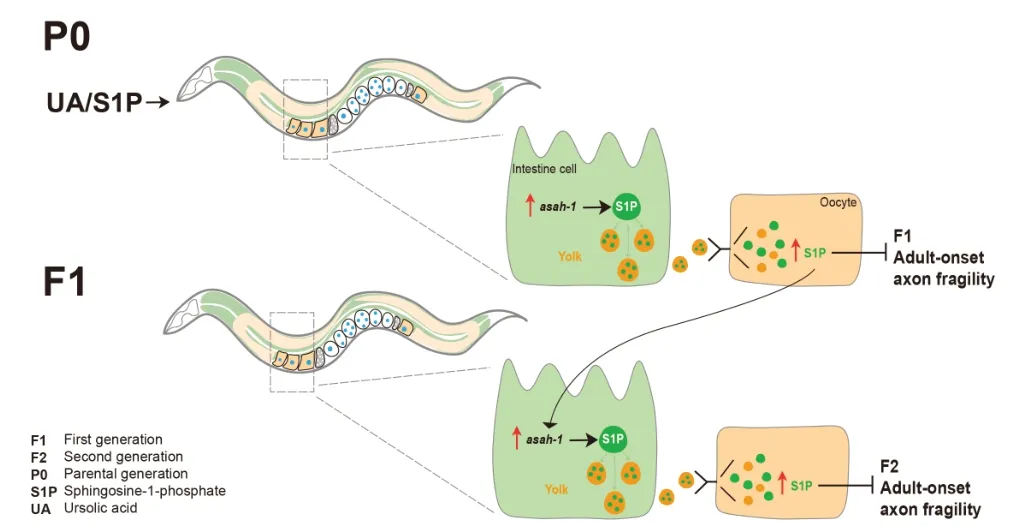
Figure 2|Proposed model of UA/S1P intergenerational neuroprotection.
Conclusion and perspectives:Overall,we showed that UA supplementation in the maternal diet can alter the provisioning of S1P from mother to offspring and prevent axon fragility of offspring intergenerationally.Our results reveal that this intergenerational neuroprotective effect is dependent on intestinal-generated S1P transport in the yolk to oocytes.Future work could focus on how altered sphingolipid homeostasis in early life affects neuronal health in aging and potential pathways that may be responsible for such modulation.Ultimately,we revealed a novel role for diet-mediated intestinal sphingolipid metabolism in neuronal health and pointed to broader underlying roles of metabolic gene expression in controlling intergenerational effects.Consistent with this,the link between maternal metabolic status and long-term changes in metabolism and health status in offspring is also observed in mammals,and our findings may provide a novel epigenetic mechanism in response to prior environmental exposure and adult disease risk in offspring.
This work was supported by the National Health and Medical Research Council grants GNT1105374,GNT1137645 and GNT2000766(to RP).
Wenyue Wang*,Roger Pocock*
Development and Stem Cells Program,Monash Biomedicine Discovery Institute and Department of Anatomy and Developmental Biology,Monash University,Melbourne,VIC,Australia
*Correspondence to:Roger Pocock,PhD,roger.pocock@monash.edu;Wenyue Wang,PhD,wenyue.wang1@monash.edu.
https://orcid.org/0000-0002-5515-3608(Roger Pocock)
Date of submission:September 21,2023
Date of decision:November 2,2023
Date of acceptance:November 17,2023
Date of web publication:December 21,2023
https://doi.org/10.4103/1673-5374.391333
How to cite this article:Wang W,Pocock R(2024)A sphingolipid message promotes neuronal health across generations.Neural Regen Res 19(11):2325-2326.
Open access statement:This is an open access journal,and articles are distributed under the terms of the Creative Commons AttributionNonCommercial-ShareAlike 4.0 License,which allows others to remix,tweak,and build upon the work non-commercially,as long as appropriate credit is given and the new creations are licensed under the identical terms.
- 中國神經再生研究(英文版)的其它文章
- Krüppel-like factor 2 (KLF2),a potential target for neuroregeneration
- Defined hydrogels for spinal cord organoids: challenges and potential applications
- Neuronal trafficking as a key to functional recovery in immunemediated neuropathies
- Advancements in personalized stem cell models for aging-related neurodegenerative disorders
- New insights into astrocyte diversity from the lens of transcriptional regulation and their implications for neurodegenerative disease treatments
- Harnessing endothelial cells and vascularization strategies for nerve regeneration